

A SPECT scan is a nuclear medicine examination that helps doctors assess how specific organs and tissues are functioning, rather than showing only their structure. It is used in several clinical areas, including heart, bone, and brain spect imaging, when functional information can support decisions about diagnosis or follow-up.
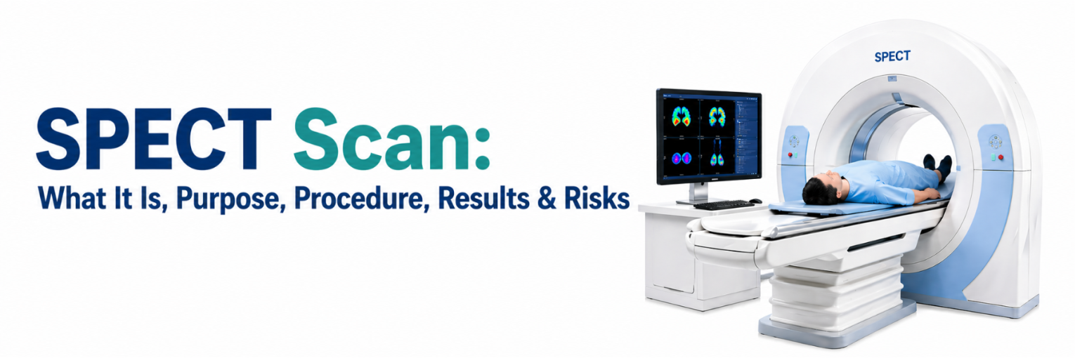
What is a SPECT scan?
In Medical imaging, SPECT stands for Single Photon Emission Computed Tomography. A SPECT scan uses a small amount of a radioactive tracer and a specialised camera to create 3D images that reflect processes such as blood flow or tissue activity.
Unlike CT or MRI, which primarily show anatomy, a SPECT scan is designed to help visualise function. This difference can matter when symptoms and routine tests do not fully explain a patient’s condition.
How Does SPECT Work?
A SPECT scan begins with a tracer that is usually injected into a vein, and for some studies, may be swallowed or inhaled. The tracer accumulates in targeted tissues in a way that relates to the process being studied, such as the regional blood supply.
Image formation typically involves:
- The tracer emits gamma rays.
- A gamma camera rotates around the body and detects these emissions.
- A computer reconstructs the signals into cross-sectional slices and 3D images.
Because uptake varies by organ system, the tracer and timing are selected for the specific study, such as cardiac perfusion, skeletal evaluation, or neurological assessment.
SPECT vs. PET Scan
The comparison of SPECT vs PET is important because both techniques are functional nuclear medicine tests, but they use different tracers and detection systems. PET uses positron-emitting tracers, while SPECT uses gamma-emitting tracers.
A doctor may consider one over the other based on:
- The clinical indication and the tracer suited to it
- Availability of equipment and radiotracers in India
- Whether combined imaging is needed, such as SPECT/CT or PET/CT
- Patient factors, including mobility, breathlessness, or difficulty remaining still
In many centres, SPECT is routinely available and is used for a broad range of indications.
Purpose of a SPECT scan
The purpose of a SPECT scan is to help assess organ system function and identify patterns that may be consistent with disease, recovery, or variation from expected findings. A doctor may request this test when it can add information for diagnosis, risk assessment, treatment planning, or monitoring.
Common SPECT scan uses include:
- Cardiac perfusion studies to evaluate blood flow to the heart muscle during rest and stress
- Bone scans to identify areas of increased bone turnover that may relate to injury, infection, or other conditions
- Selected neurological studies, including brain SPECT imaging, to assess regional perfusion patterns
- Evaluation of certain endocrine and inflammatory problems when an appropriate tracer is indicated
A SPECT scan is interpreted alongside history, examination, blood tests, and other imaging. Findings may prompt correlation with additional investigations rather than serving as a single deciding test.
The SPECT scan Procedure
Your experience during a spect procedure usually follows a planned sequence, although the timing varies depending on the type of study. In India, these scans are commonly performed in a nuclear medicine department with trained technologists and a nuclear medicine physician.
Before the scan
SPECT preparation depends on whether the study is cardiac, bone, or neurological. Inform the team about your medicines, allergies, and any recent imaging.
Preparation may include:
- Carrying prior reports, prescriptions, ECGs, and earlier scan images if available
- Following food and drink instructions, which may include fasting for a specified period for some studies
- Avoiding caffeine for a cardiac perfusion study when advised, including coffee, tea, cola, energy drinks, and some cold or pain medicines
- Discussing diabetes medicines, if fasting is required, so that timing can be adjusted safely
You may be asked to remove jewellery and other metal items and to wear comfortable clothing. For treadmill-based stress testing, suitable footwear may be requested.
At registration, you may be asked to provide identification and to complete a short questionnaire about pregnancy, breastfeeding, and recent procedures. If you have had a recent contrast CT, angiography, or surgery, share dates, as this can affect scheduling and interpretation. For cardiac studies, the team may review whether any medicines should be held; do not stop medicines unless your doctor advises it.
During the scan
After tracer administration, there is often a waiting period to allow uptake in the target tissue. The scan is performed while you lie on an imaging table as the camera rotates around you.
During scanning, you may notice:
- Positioning supports to help you remain still
- A scan time that may be divided into more than one set of images
- Extra CT images in some centres when SPECT/CT is used to improve localisation and reduce artefacts
The camera can get close to your body to improve signal quality, but it does not fully enclose you. If you feel anxious, tell the staff early so they can guide you.
After the scan
Most people can return to routine activities unless specific instructions are given. Depending on the tracer and study, you may be advised to drink fluids to help eliminate tracer material through urine.
Understanding SPECT scan Results
SPECT scan results are reviewed by a nuclear medicine physician and reported with the referral question in mind. Reports typically describe tracer distribution, whether uptake looks within expected limits, and whether there are areas of relative increase or decrease.
Findings may be described as:
- Typical distribution for the protocol used
- Reduced uptake areas, which may suggest reduced perfusion or function
- Increased uptake areas, which may reflect higher activity, inflammation, or healing, depending on the organ studied
- Artefacts related to movement, soft tissue attenuation, or technical factors
Reports are often available the same day or within 24-48 hours, depending on the centre’s workflow. Keep a copy for future comparison, particularly if repeat imaging is being considered.
For cardiac perfusion, reports may comment on differences between rest and stress images and may include quantitative scores in some centres. For bone studies, focal uptake is described by location and intensity. For neurological studies, patterns in brain SPECT imaging are interpreted cautiously and are combined with clinical assessment and other tests.
Risks and Safety of SPECT Scans
Overall spect safety considerations include tracer-related radiation exposure, patient-specific precautions, and uncommon side effects. The nuclear medicine team aims to keep exposure as low as reasonably achievable while obtaining images that answer the clinical question.
Radiation Exposure in SPECT Scans
A SPECT scan involves ionising radiation from the tracer, and sometimes from a low-dose CT component if SPECT/CT is performed. The amount depends on the tracer, dose, and protocol.
Points often discussed include:
- Doses follow established protocols and may be adjusted for patient factors
- Exposure varies between study types, so comparisons should be study-specific
- The expected medical value of the information is weighed against radiation exposure for each patient
If you have concerns, ask why the test is advised and whether alternatives are suitable for your situation.
Who Should Avoid SPECT Scans?
A SPECT scan may be deferred or modified in situations where radiation exposure should be minimised, or the protocol is not suitable.
Tell your doctor and the imaging team if you:
- Are pregnant or think you may be pregnant
- Are breastfeeding, as a temporary pause, may be advised depending on the tracer
- Have had another nuclear medicine scan recently
- Cannot lie flat or remain still due to pain, breathlessness, or severe anxiety
The team may adjust timing, support positioning, or discuss alternatives based on clinical priorities.
Side Effects of SPECT Scans
Side effects are uncommon and are usually related to the injection or, in cardiac studies, the stress component.
Possible issues include:
- Mild pain, bruising, or swelling at the injection site
- Short-lived dizziness, headache, or nausea with some tracers or stress medicines
- Rare allergic-type reactions, which are managed by trained staff when they occur
- Discomfort from having to stay still during image acquisition
If you feel unwell during or after the test, inform the staff promptly.
Conclusion
A SPECT scan can provide functional information that complements structural tests and supports clinical decision-making when a specific question needs clarification. When the study is advised, the referral reason, the chosen protocol, and the interpretation process matter as much as the images themselves.
If you are scheduled for a scan, follow the instructions for SPECT preparation, share a complete medicine list, and disclose pregnancy or breastfeeding status before the tracer is given. Discussing concerns in advance can help the team plan the study and reduce the chance of avoidable repeat imaging.
FAQs
How Long Does a SPECT scan Take?
Total visit time can range from one to several hours because tracer uptake often requires waiting. The camera-scanning portion may take 20 to 60 minutes, depending on the protocol and whether images are acquired across multiple phases.
Is a SPECT scan Painful?
The scan is not usually painful. You may feel a brief sting during the injection and mild discomfort from lying still. If you have back or joint pain, inform the staff so that positioning support can be arranged.
How Should I Prepare for a SPECT scan?
Follow the instructions provided by your centre, as preparation varies by study type. You will usually be asked about medicines, allergies, pregnancy, breastfeeding, and recent scans. Some protocols require fasting or avoiding caffeine, and cardiac studies may include guidance about exercise and medicines.
When is a SPECT scan Recommended?
A doctor may recommend the test when functional information is needed to assess blood flow, evaluate bone activity, or support treatment planning and monitoring. The decision depends on symptoms, examination findings, and the question being addressed.
Can a SPECT scan show brain damage?
In some settings, brain SPECT imaging may show perfusion patterns that can be associated with neurological conditions. However, it does not directly demonstrate tissue injury in the way structural imaging can, and results require specialist interpretation with clinical assessment and other tests.
What are the benefits of SPECT?
Benefits may include the ability to evaluate function and identify patterns not visible on structural scans, when interpreted alongside other clinical information. In many Indian centres, SPECT is also more accessible than some advanced functional imaging options.
Is SPECT scan dangerous?
A SPECT scan involves radiation exposure, and the test is planned so that the expected medical benefit outweighs potential risk. Side effects are uncommon, and additional precautions are used for pregnancy, breastfeeding, and patient-specific needs. If you remain concerned, discuss this with your doctor before the appointment.