

Urinary symptoms can feel disruptive, especially when they recur or when there is concern about an injury. In such situations, your clinician may advise an imaging test that looks specifically at the urinary bladder. Cystography is designed for that purpose: it outlines the bladder using a contrast agent and produces images that can be reviewed by a radiologist and your treating doctor.
This article explains what the test involves, why it may be advised, how it is carried out, and what preparation usually includes.
Decisions about whether the test is suitable should always be made with the opinion of a qualified clinician who knows your symptoms and medical history. If you feel anxious, ask the team to explain each step and expected sensations beforehand.
What is Cystography?
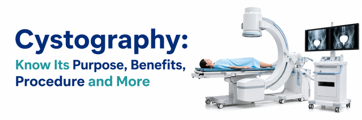
Cystography definition: Cystography is a radiology examination in which the bladder is filled with a contrast medium through a urinary catheter and then imaged, most commonly with X-ray and fluoroscopy. The images obtained are often referred to as a bladder cystogram, because the contrast outlines the inner surface of the bladder and allows the radiologist to assess its shape, position, and integrity.
In day-to-day practice, a clinician may use the word cystography to describe more than one related study. The main formats include:
- A contrast cystogram, where images are taken while the bladder is filling with contrast
- A voiding study, where images are captured as urine is passed, is often called a voiding cystourethrogram
- A CT cystogram, where contrast is instilled into the bladder and CT images are acquired, is usually performed when a detailed evaluation for injury is required
How the images are interpreted
During filling, the radiologist looks for a smooth, continuous contrast outline and checks whether contrast remains within the bladder. A focal outpouching may be described as a diverticulum, while an irregular contour can prompt correlation with clinical history and other imaging. If contrast is seen outside the expected bladder boundary, it may suggest extravasation and is reported with details on location and extent. In a voiding study, attention also includes whether contrast tracks up towards the kidneys, which can indicate reflux, and whether the urethral passage appears narrowed or disrupted during voiding.
Because the examination is tailored to a question, it is usually ordered with a specific suspected diagnosis in mind. Findings are therefore interpreted together with urine tests, symptoms, and prior scans, and your doctor may recommend additional evaluation if results do not fully explain the complaint.
The test is performed in a controlled radiology setting. A radiographer and radiologist supervise imaging, and a nurse or trained staff member assists with patient preparation, catheter placement, and infection prevention measures. The final report is interpreted alongside your symptoms, urine tests, and other imaging, because the study is intended to answer a specific clinical question rather than serve as a general screening test.
Need for Cystography
A cystography test may be advised when a doctor needs visual information about the bladder that cannot be obtained reliably from a standard X-ray or ultrasound alone. In cystogram radiology, contrast within the bladder acts as an internal outline, making it easier to assess the bladder wall contour and to look for leakage of contrast outside the bladder.
When the test may be recommended
The decision to perform the test depends on symptoms and clinical findings. Your doctor may consider it in situations such as:
- Suspected bladder injury after pelvic trauma, a fall, or a road traffic accident
- Concern about a urine leak after pelvic surgery, catheter-related complications, or certain urological procedures
- Recurrent urinary tract infections when vesicoureteral reflux needs assessment, particularly in selected paediatric cases
- Assessment of structural conditions, such as bladder diverticula, requires an outline of the bladder cavity
- Evaluation of certain voiding problems when a voiding study is requested to visualise the urethra during urination
What a bladder cystogram can show
A contrast study may help the radiologist evaluate:
- The general shape and capacity pattern of the bladder during filling
- Irregularities along the bladder outline that may suggest structural change
- Leakage of contrast that may indicate a tear or perforation
- Reflux of contrast into one or both ureters during voiding studies
- Urethral appearance during voiding studies, which can be relevant in selected cases
It is equally important to understand what the test does not do. It does not assess kidney function, and it does not replace urine tests for infection. Your clinician may advise other investigations, such as ultrasound, CT, MRI, cystoscopy, or laboratory tests, depending on the suspected condition.
The test is not a routine screening examination. It is generally planned after your clinician reviews symptoms and decides that contrast imaging may add information beyond ultrasound or plain radiography in your case.
Benefits of Cystography
The potential benefits of Cystography relate to the accuracy of answering a targeted question about the bladder. Depending on the indication, the test may offer the following advantages:
- A clear contrast outline that can make urine leakage easier to recognise
- Dynamic assessment with fluoroscopy, allowing observation during filling and, when requested, during voiding
- Support for clinical decision-making after trauma or surgery when bladder integrity is being evaluated
- Imaging that is available in many centres and can usually be completed within a single visit
- A structured report that can be compared with follow-up studies when clinically indicated
Cystography Test Procedure
The cystogram procedure is planned around the clinical question, so the steps may vary slightly between a standard study, a voiding study, and CT-based imaging. Your care team should explain the purpose of the test, what you may feel during the examination, and how the results will be used before the examination begins.
Before the test: preparation and safety checks
Most centres perform basic checks to reduce avoidable risks. These commonly include confirming your identity, reviewing allergies, and asking about current medicines. If there are symptoms of a urinary infection, your clinician may decide whether treatment is required before imaging.
Preparation instructions can differ between centres and between X-ray and CT protocols. CT cystogram preparation may involve additional steps because timing and scan phases can affect image quality. For many patients, the preparation for a standard contrast study may include:
- Informing the team about any prior contrast reaction, asthma, or severe allergies
- Sharing details of kidney disease, bleeding disorders, recent urinary procedures, or catheter problems
- Telling the team if pregnancy is possible, as radiation exposure requires careful risk assessment
- Bringing relevant reports such as ultrasound findings, urine culture results, discharge summaries, or operative notes
- Wearing comfortable clothing and following any guidance about eating, drinking, or arriving with an empty bladder
If a child is undergoing a voiding study, the approach may be adapted for comfort and cooperation. Parents or guardians are often counselled about how the catheter is placed and how images are obtained. Some centres may use distraction techniques; sedation is not routine and, if considered, is decided by the treating team.
During the test: what happens in the imaging room
Once you are on the imaging table, the genital area is cleaned with an antiseptic to reduce infection risk. A thin sterile catheter is inserted through the urethra into the bladder. This step can cause stinging or pressure, and the intensity of discomfort varies from person to person.
After catheter placement, contrast is introduced slowly. The radiology team monitors filling and captures images from different angles. Fluoroscopy may be used to observe contrast movement in real time. You may be asked to change position, take a breath, or hold still for a short period while images are recorded.
If the study requested is a voiding examination, you may be asked to pass urine while images are taken. This portion is often performed with privacy measures in place, but it can still feel awkward. The team is trained to conduct the process professionally and to minimise discomfort while obtaining the required diagnostic views.
After the test: immediate care and what you might notice
When imaging is complete, the catheter is removed unless a clinician advises otherwise. You may be encouraged to drink fluids and pass urine to help clear contrast from the bladder. Mild burning during urination, increased urge to pass urine, or a small amount of blood on wiping can occur temporarily due to catheter-related irritation.
In most cases, the discomfort settles with hydration and observation. However, you should seek medical advice if you develop a fever, chills, worsening lower abdominal pain, persistent burning, difficulty passing urine, or significant blood in your urine, as these may indicate complications such as infection or urinary retention.
Risks and side effects to discuss with your doctor
Like any invasive imaging test, cystography involves specific risks. The likelihood and significance depend on your health status and the reason the test is being done. Points that are commonly discussed include:
- Radiation exposure from X-ray or CT imaging, which is usually minimised using standard protection principles
- Urinary tract infection risk related to catheterisation, particularly in patients with prior infections or urinary abnormalities
- Temporary discomfort, burning, or mild bleeding after catheter removal
- Rare contrast reactions; risk assessment is important if there is a history of allergy
Your clinician may advise whether any medicines should be paused or whether antibiotics are required. Antibiotics are not automatically needed for every patient and should be used only when prescribed.
Understanding the report and next steps
The radiologist evaluates images for the requested findings, such as contrast leak, reflux, or abnormal bladder contour. The report may also note technical aspects, including whether filling was adequate and whether voiding images were obtained. Your treating doctor then correlates the report with symptoms and other test results to decide on further management.
Cystogram procedure steps
Although details can vary, the essential steps of a cystogram are usually: sterile catheter placement, controlled instillation of contrast, imaging during filling, and catheter removal, with additional images during voiding when a voiding study is requested. If a CT cystogram is planned, the CT is performed after the bladder is filled, and the protocol is adjusted based on the suspected injury pattern.
Cystography at Vijaya Diagnostics
When your doctor advises Cystography, the quality of preparation, infection control, and communication can influence how smoothly the appointment goes. At Vijaya Diagnostics, patients are generally guided through appointment scheduling, pre-test instructions, and supervised imaging in a dedicated radiology area, with attention to privacy and radiation protection practices.
For information on availability, centre locations, and booking, refer to the Vijaya Diagnostics home page and select the service route that matches your clinician’s request, such as a standard study or a voiding study. Your treating doctor will decide whether the test is appropriate for you and how the findings fit into your overall treatment plan.
FAQs
What is the primary purpose of a Cystography procedure?
The primary purpose of Cystography is to create a contrast outline of the bladder so that the radiologist can assess bladder integrity, contour, and, when relevant, urine leak or reflux.
How does a Cystography differ from a standard X-ray?
A standard X-ray relies on natural differences in tissue density and does not outline the bladder cavity well. In Cystography, contrast inside the bladder provides a visible boundary, which can make certain findings clearer than a plain film.
Is the procedure painful for the patient?
The most uncomfortable part is often catheter insertion and the sensation of bladder filling. Many people report pressure or burning rather than severe pain, but discomfort levels can vary and should be discussed with the team beforehand.
What are the main benefits of choosing Cystography?
When appropriately indicated, Cystography may provide focused information about bladder injury, contrast leak, reflux, or structural change, supporting treatment decisions that depend on bladder imaging.
What is a “Voiding Cystourethrogram” (VCUG)?
A voiding cystourethrogram is a type of cystography in which images are taken while the patient passes urine. It is often used to evaluate reflux and to visualise the urethra during voiding.
What are the risks or side effects associated with the test?
Possible risks include urinary infection, short-term burning with urination, mild bleeding, and radiation exposure. Contrast reactions are uncommon, but it is important to inform the team of any prior reactions to contrast or severe allergies.
How should a patient prepare for a Cystography?
Preparation usually includes sharing medical history, medicines, allergy details, and pregnancy status, and following centre-specific instructions about arrival time, hydration, and bladder emptying. If you have symptoms of infection, your doctor may advise whether urine testing or treatment is needed before the test.