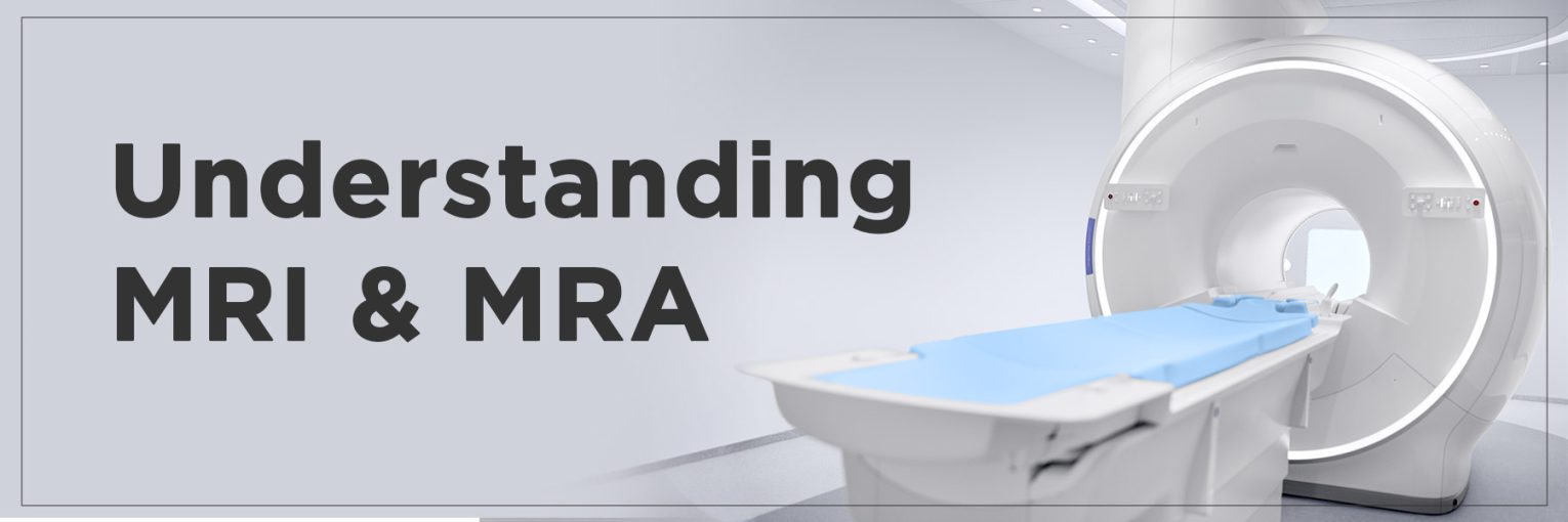
MRI and MRA are powerful imaging tools that help doctors diagnose health issues with precision. MRI, or magnetic resonance imaging, creates detailed pictures of organs, tissues, and bones using strong magnets and radio waves. It’s ideal for spotting brain tumours or joint injuries. MRA, a specialised MRI, zooms in on blood vessels to detect blockages or aneurysms.
Both are non-invasive and radiation-free, offering clear insights safely. The 1.5T MRI, with its balanced clarity and speed, is widely used. This post explores how they work MRI MRA, their differences, and when each is the best choice for your health.
What is MRI?
Magnetic resonance imaging, or MRI, is a medical technique that uses a strong magnetic field and radio waves to create detailed pictures of soft tissues and organs. During an MRI scan, you lie inside a large, tube-shaped magnet, and the MRI process aligns hydrogen atoms in your body to produce clear cross-sectional or 3D images.
A 1.5T MRI, with its 1.5-tesla magnet, offers sharp images and is widely used for its balance of quality and speed. MRI benefits include being noninvasive and highly accurate, making it ideal for imaging the brain to detect tumours, strokes.
What is an MRA?
Magnetic resonance angiography (MRA) is a specific form of MRI sequence that visualises blood vessels throughout the body. The main function of MRA vs. MRI is to separate them from each other because MRA specifically examines blood vessels for conditions including aneurysms and blockages. Your position inside the large MRI tunnel enables radio waves combined with magnetic fields to create clear images.
An MRA procedure does not require invasive catheters because it remains noninvasive. The procedure requires occasional administration of contrast dye to achieve better vessel visibility. Healthcare providers use MRA for brain imaging to find weakened blood vessels and restricted blood flow in a safe manner.
MRA vs MRI: Key Differences
Here are the key differences between the two:
| Feature | MRI | MRA |
| Purpose | MRI, or magnetic resonance imaging, creates detailed pictures of soft tissues, organs, and bones. | MRA, or magnetic resonance angiography, focuses on imaging blood vessels. |
| Difference between MRI and MRA | MRI vs MRA lies in what they target—MRI scans the body’s tissues like muscles or ligaments. | MRA is a type of MRI that specifically examines blood vessels for issues. |
| Applications | MRI is used to find damage in tissues, such as torn ligaments from a fall or tumours in organs. | MRA detects blood vessel problems, like clots causing leg swelling or aneurysms. |
| When Used | Doctors order an MRI to check for injuries to soft tissues or conditions like joint damage. | MRA is chosen when blood flow issues, such as blockages or vessel damage, are suspected. |
How Does a 1.5T MRI Work?
A 1.5T MRI is a widely used MRI technology that creates clear images of the body using a magnetic field strength of 1.5 tesla. During an MRI scan, the 1.5T MRI’s magnet aligns hydrogen atoms in the body, and gradients—smaller magnets—adjust the field to capture detailed pictures. Its channel coils, like head or spine coils, improve image quality for areas like the heart or joints.
The 1.5T MRI balances good image quality with reasonable scan times, making it versatile for most studies. Compared to a 3T MRI, which has a stronger magnet and sharper images, the 1.5T is more affordable and widely available, though it may take slightly longer for some scans.
Benefits of MRI and MRA
MRI Benefits:
- Non-invasive imaging: Allows doctors to view organs, tissues, and bones without any surgery.
- Detailed pictures: Creates clear, high-quality images to detect brain abnormalities or joint injuries.
- No radiation: It uses magnetic fields and radio waves, making it safe and preventing harmful radiation exposure.
- Contrast agent use: Sometimes includes contrast to enhance the visibility of scanned areas for better diagnosis.
MRA Benefits:
- Non-invasive imaging: Examines blood vessels without the need for invasive procedures.
- Clear vascular images: Provides sharp pictures of blood flow to identify issues like strokes or heart disease.
- No radiation: Relies on magnetic fields and radio waves, ensuring safety with no radiation risks.
- Specialised focus: This speciality focuses on blood vessel conditions, such as carotid artery disease or aortic issues, to ensure an accurate diagnosis.
When to Choose MRI or MRA?
When to Choose an MRI
MRI is ideal for examining soft tissues, like the brain or spinal cord, to find issues not related to blood vessels.
- Brain tumours: MRI detects abnormal growths in the brain, helping doctors plan treatment.
- Multiple sclerosis: It shows damage to nerve coverings caused by this immune system disorder.
- Spinal cord injuries: MRI reveals damage to vertebrae or ligaments from accidents.
- Inner ear or eye disorders: It diagnoses conditions like inflammation causing dizziness.
- Traumatic brain injury: MRI spots brain damage from blows or penetrating objects.
When to Choose an MRA?
- Why You Might Need an MRA: MRA is used to check blood vessels for problems like blockages or abnormal growths.
- Strokes: It identifies reduced blood flow to the brain from narrowed arteries.
- Aneurysms: MRA finds weak spots in blood vessels, like in the brain or aorta.
- Carotid artery disease: It detects plaque buildup that could lead to stroke.
- Kidney or leg artery issues: MRA evaluates narrowed vessels before surgery or stenting.
- Arteriovenous malformations: They reveal abnormal vessel connections in the brain or body.
FAQs
What is the difference between MRI and MRA?
A device known as an MRI uses magnets combined with radio waves to produce images of body organs, muscles, and bones. Doctors rely on MRI to detect both skeletal and neurological problems. MRA represents magnetic resonance angiography, which functions as a specific MRI technique designed to examine only blood vessels. Medical professionals use this method to detect blocked arteries in patients. MRI delivers images of tissue structure, while MRA shows images of vascular structures.
Is MRA better than MRI?
MRA isn’t better than MRI, and MRI isn’t better than MRA. They do different jobs. If a doctor wants to see your muscles or brain, they pick an MRI. If they need to check your blood vessels for something like a blockage, they choose MRA. It depends on what part of the body they’re worried about.
How long does a 1.5T MRI take?
A 1.5T MRI scan usually lasts between 20 and 30 minutes. It depends on what the doctor is looking at. For example, scanning your knee might be faster than scanning your whole back. You lie inside the machine, and it takes pictures. If the doctor uses a special dye to make things clearer, it could take a bit longer.
Is an MRA scan painful?
No, an MRA scan doesn’t hurt. You just lie inside a machine that takes pictures of your blood vessels using magnets and radio waves. There’s no cutting or poking. Sometimes, a dye is injected to help see the vessels better, and you might feel a small pinch from the needle, but that’s it.
Can MRI and MRA detect cancer?
Yes, an MRI can show if there’s a tumour in places like your brain or liver, which might be cancer. It gives clear pictures of tissues. MRA can check if a tumour is affecting nearby blood vessels. Both help doctors figure out if cancer is there, but they might need to do other tests, like taking a small sample, to be sure.
MRI Scan Centre
To find MRI Scan Centre near me click here.


