

A CT temporal bone scan is a special type of imaging study used to obtain detailed cross-sectional images of the bones that surround the ear. This scan is routinely ordered when a patient is suffering from hearing loss, earache, or following trauma to the side of the head. It produces high-resolution images to help doctors identify conditions that are otherwise hard to spot with ordinary X-rays. Rapid, non-invasive, and extremely accurate, computed tomography of the temporal bone can often be a vital step to evaluate many disorders that affect the ears and skull.
What is CT Temporal Bone Scan?
A CT temporal bone scan is a special type of diagnostic imaging that provides detailed images of the bones around the ears on both sides of the skull. With the help of X-rays and computer processing, it produces cross-sectional images that enable doctors to scrutinize the tiny and complex structures inside and around the ear. This refers to the ear canal, the bones in the middle ear, the inner ear, and the surrounding tissues.
A CT temporal bone scan is frequently advised if there is unexplained hearing loss, persistent ear infections, or head trauma. It can also help in surgical mapping prior to ear surgeries.
The CT temporal bone scan is safe, painless, and requires just a few minutes. It’s a more exact form of imaging than standard X-rays, and it can detect nuanced issues like minuscule bone fractures or concealed infections. An accurate diagnosis and effective treatment planning require the clarity and detail of a temporal bone CT scan.
Purpose of a CT Temporal Bone Scan
The temporal bone scan purpose in imaging is to show if there is anything peculiar about the ears and hyperdense and low-density bones surrounding them. There are multiple justifications for ordering a CT temporal bone scan.
- One of the most common is to identify fractures caused by trauma. The scan gives a detailed view of the delicate bones in the ear and skull base that are often too small to visualize with conventional imaging.
- Another common use is to identify infections like mastoiditis, inflammation of the mastoid bone that occurs when untreated ear infections spread.
- A temporal bone CT scan protocol standardizes the scan process to help maintain consistency in the quality of care and diagnosis.
- It is also crucial in diagnosing congenital abnormalities — medical conditions present at birth that affect hearing or the structure of the ear.
- The scan may also help plan surgical interventions, such as cochlear implants or the removal of tumors.
Experts utilize CT temporal bone filming to investigate the intricate internal structure, such as the cochlea and semicircular canals. The CT scan serves as a guide to comprehend, diagnose, and treat numerous ear-related issues effectively.
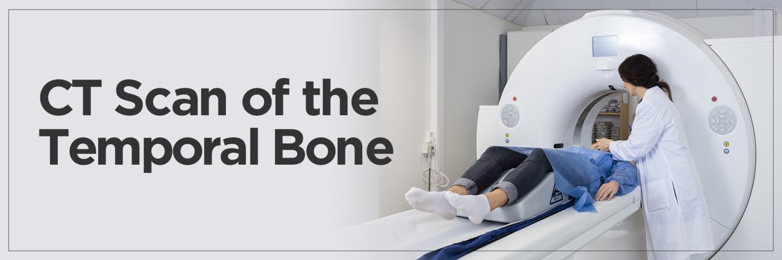
Procedure: How is a CT Temporal Bone Performed?
The process of a CT Temporal Bone scan is smooth and typically takes less than 30 minutes.
- Before the scan begins, the patient may need to remove jewelry or metal objects that could interfere with image clarity.
- There’s usually no need for fasting or special preparation unless contrast dye is being used, which is rare for temporal bone studies.
- Once ready, the patient lies on a narrow table that slides into the CT machine.
- For precise temporal bone CT scan planning, the head is gently secured to avoid any movement. The scan is painless, but remaining still is important to produce sharp, accurate images.
- During the scan, the machine takes multiple cross-sectional X-rays, which are reconstructed by a computer into detailed 3D images of the temporal bones.
Understanding temporal bone CT anatomy is crucial for radiologists, as the scan captures minute structures, including the ossicles (tiny ear bones), ear canal, and inner ear components.
The CT temporal bone filming protocol ensures each scan is performed consistently, while the temporal bone CT scan protocol helps align the angles and positions to best view the anatomy.
Once completed, the images are reviewed by a radiologist, and the results are shared with the referring doctor.
Risks and Considerations
Temporal bone CT scan planning is a critical step that aims to optimize image clarity while minimizing radiation exposure. Modern CT machines and refined scanning protocols have significantly reduced the radiation dose needed, making the scan safer than ever before. Still, it’s important for patients to discuss any health concerns, prior scans, or allergies with their doctor to ensure a tailored and safe imaging approach.
- While a CT temporal bone scan is generally considered safe and non-invasive, it does involve exposure to a small amount of ionizing radiation. For most adults, this level of radiation is minimal and poses little long-term risk.
- Repeated scans or scans performed on younger patients, such as children, should be carefully justified, as developing tissues are more sensitive to radiation. Therefore, physicians usually weigh the diagnostic benefits against potential risks before recommending the scan.
- In cases where contrast dye is needed, though rare for temporal bone imaging, some individuals may experience allergic reactions or kidney-related side effects, particularly those with pre-existing kidney conditions.
- Pregnant women are another group for whom the scan is typically avoided unless absolutely necessary, due to the potential effects of radiation on the developing fetus.
CT Temporal Bone vs. MRI: Which is Appropriate?
When it comes to evaluating conditions affecting the ear and skull base, CT and MRI offer powerful imaging capabilities, but each has distinct advantages.
- A CT temporal bone scan excels in capturing detailed images of bony structures. It is ideal for diagnosing fractures, chronic ear infections, or congenital abnormalities where fine bone detail is crucial. The high-resolution imaging makes it particularly useful for assessing ossicles, cochlear structure, and the bony labyrinth.
- On the other hand, MRI is superior in evaluating soft tissue structures. It is the preferred choice for detecting nerve-related issues, inner ear inflammation, tumors such as acoustic neuromas, or vascular abnormalities. Unlike CT, MRI does not make use of ionizing radiation, which makes it safer for repeated imaging or use in pregnant patients—though it is typically more time-consuming and costly.
Choosing between the two often depends on what the physician is looking to assess. Temporal bone CT scan planning helps ensure the scan focuses on bone details with minimal exposure, whereas MRI planning centers around highlighting nerve and soft tissue anatomy. In some complex cases, doctors may even recommend both scans to provide a complete picture of the issue.
FAQs
1. What are the symptoms of a temporal bone problem?
Temporal bone issues can present a variety of symptoms depending on the specific condition. Common signs include persistent ear pain, hearing loss, ringing in the ears (tinnitus), dizziness or vertigo, discharge from the ear, and sometimes facial weakness or paralysis. Infections or injuries to the temporal bone can also cause swelling, tenderness around the ear, or even balance disturbances due to inner ear involvement.
If you experience any of these symptoms, a healthcare provider might recommend imaging, such as a CT temporal bone scan, to investigate further.
2. What does a CT bone scan show?
A CT bone scan—specifically a CT of the temporal bone—produces highly detailed images of the ear’s bony structures, including the ear canal, middle and inner ear, and surrounding skull base. It can detect fractures, infections like mastoiditis, congenital malformations, and even bone erosion from chronic diseases.
The scan can also assist doctors in surgical planning by mapping the anatomy with precision. It offers cross-sectional and 3D views, helping clinicians diagnose conditions that are not visible through physical examination alone.
3. Why is the temporal bone important?
The temporal bone is very important for both hearing and balance. It houses the external auditory canal, middle ear bones (ossicles), cochlea, and semicircular canals—all vital structures for sound transmission and spatial orientation. It also provides protection for the facial nerve and major blood vessels.
Damage or disease affecting the temporal bone can significantly impair a person’s quality of life, affecting not just hearing but also facial movement and balance. That’s why timely diagnosis using tools like a CT scan is so important for early treatment and management.
4. What would damage to the temporal bone cause?
Damage to the temporal bone—whether from trauma, infection, or chronic disease—can have a range of consequences. It may lead to conductive or sensorineural hearing loss, facial nerve palsy, chronic ear infections, vertigo, or even leakage of cerebrospinal fluid in severe cases.
Some people might also experience difficulty in speech recognition or sensitivity to loud sounds. Identifying the extent and exact spot of the damage often requires a CT temporal bone scan for accurate treatment planning.
5. What is the difference between CT head and CT temporal bone?
A CT head scan provides a broad overview of the brain, skull, sinuses, and surrounding structures, useful for detecting strokes, bleeding, tumors, or trauma. However, it lacks the fine resolution needed to examine ear structures in detail. In contrast, a CT temporal bone scan is specifically focused and high-resolution, designed to capture the intricate bony anatomy of the ear and nearby areas.
While both use similar technology, CT temporal bone scans use specialized settings and positioning for maximum clarity of the ear region.