

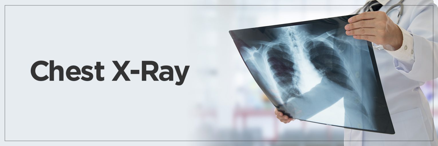
A chest x-ray is one of the most frequent diagnostic tests and assists doctors in obtaining a fast view of the organs and structures inside the chest. It is known as an X-ray Chest AP View when it is taken from the front to back. This chest X-ray is particularly helpful for patients in emergency or critical care.
It provides crucial details about the lungs, heart, airways, and bones, helping doctors make prompt and accurate decisions. Let’s explore this view, how to implement it, and why it matters.
What is an X-ray Chest AP View?
So an X-ray Chest AP View will be taken from the front of the body to the back side. It is a very useful chest X-ray. “AP” means anteroposterior, indicating that the X-ray beam travels from front (anterior) to back (posterior) of the chest. This is often employed with patients who are unable to sit erect, such as those who are bedridden or in the emergency department. It provides a rapid way for doctors to evaluate the heart, lungs and surrounding structures.
A normal chest X-ray would show well-defined lung fields, a clearly defined heart, and visible bones. But, an X-ray Chest AP View sometimes shows the size of the heart as slightly enlarged, which doctors take into consideration when diagnosing. This vantage point is an effective one when other X-ray positions are not an option.
What is an AP View Chest X-Ray?
In a case where a person can’t stand, an AP view chest X-ray is done with the patient in a lying or sitting position. The X-ray machine is positioned in front of the chest, and the film or detector is located behind your back.
An Anteroposterior chest X-ray is particularly helpful in critical care for young or old patients. It very much differs from a standard PA (posteroanterior) view, in which the image is projected from back to front and is usually more reliable for estimating the size of the heart.
Lateral views provide side-on views, however. So, AP view takes less time to position the patient, which is beneficial for bedside imaging when it comes to chest X-ray positioning. If someone cannot get low enough, this view can be a bit less detailed than other views, but it is still an important imaging technique when a patient is immobilised.
Why is an AP View Chest X-Ray Done?
An AP view chest x-ray is often used in certain medical scenarios when alternative imaging positions are not possible or practical. One example is that it is often used as an ICU chest X-ray that enables doctors to check the lungs and heart of critically ill patients without moving them.
It also acts as a portable chest X-ray, which is usually performed at the patient’s bedside with a mobile X-ray unit. That kind of flexibility is vital in emergency rooms and operating theatres.
In addition, a supine chest X-ray (patient is flat) is another type of AP view chest X-ray performed on patients who are unable to sit. These situations render the AP view an important imaging test in recognition of ailments such as pneumonia, heart failure, pleural effusion, and the placement of clinical equipment such as tubes and catheters.
How is the AP View Chest X-Ray Performed?
The chest X-ray procedure in an AP (anteroposterior) view is typically performed when the patient is unable to stand or sit upright, such as in emergency rooms, ICUs, or during surgical recovery.
- In this setup, the patient either sits upright in bed or lies flat on their back.
- The X-ray machine is placed in front of the chest, and the film or digital detector is positioned behind the patient’s back.
- Proper chest X-ray positioning is crucial to obtaining a useful image. The patient is usually asked to stay still and hold their breath briefly to prevent motion blur.
- The X-ray beam travels from front to back (anterior to posterior), capturing the internal chest structures.
- Since the AP view is often done with portable equipment, technicians take care to align the machine correctly to avoid skewed or distorted images.
- Safety measures are always followed to minimise radiation exposure. Protective gear such as lead aprons may be used, and only the necessary body part is exposed to the X-ray.
This careful and quick process ensures patients get the imaging they need with minimal discomfort or movement.
What Can an AP Chest X-Ray Detect?
An AP chest X-ray can reveal a wide range of chest X-ray abnormalities that help doctors diagnose and treat various conditions.
- One of the most common findings is pneumonia on X-ray, which appears as localised or diffuse areas of increased opacity in the lungs, indicating infection or inflammation.
- In cases of fluid buildup around the lungs, a condition called pleural effusion, the X-ray shows fluid levels that might shift with changes in position.
- It can also help identify chest X-ray heart size abnormalities, such as cardiomegaly, which is an enlarged heart. However, due to the beam direction, the AP view may slightly exaggerate heart size.
- Other possible findings include lung masses or tumours, which appear as shadowy lesions, as well as signs of chronic lung diseases like COPD or emphysema.
- The AP view is also useful for checking the placement of medical devices, such as pacemakers, endotracheal tubes, and central lines.
Despite some limitations, this view remains a critical tool for quick diagnosis, especially in emergency and critical care settings.
AP View vs. PA View Chest X-Ray & Limitations
Understanding the differences between AP vs PA chest X-ray views is important for accurate diagnosis.
- The PA (posteroanterior) view is taken with the X-ray beam passing from back to front, typically with the patient standing and the chest pressed against the film. This position gives a clearer and more accurate view of the heart and lungs.
- In contrast, the AP view is often used as a bedside chest X-ray when patients are immobile. While convenient, this view has limitations. One major issue is magnification distortion—since the heart is farther from the film in the AP view, it can appear larger than it actually is, making it harder to assess true heart size. Additionally, the AP view can obscure or compress certain lung fields, especially at the base, which may affect the visibility of abnormalities.
While both views provide essential clinical information, the PA view is generally preferred for routine evaluations due to its higher accuracy. However, in urgent or intensive care scenarios, the AP view is invaluable for its accessibility and speed, even if the image quality may not be as detailed.
FAQs
1. Are chest X-rays risky?
Chest X-rays are generally very safe and involve a low dose of radiation. For most people, the benefits of getting a chest X-ray far outweigh the risks. The radiation exposure is minimal and not enough to cause immediate harm. However, repeated or unnecessary exposure should be avoided, especially in children and pregnant women. Medical professionals always follow safety protocols, like using lead aprons and minimising the number of images taken, to ensure your safety.
2. How does a chest X-ray work?
A chest X-ray works by sending a controlled beam of X-rays through the body, particularly the chest area. As these X-rays pass through different tissues, like the lungs, heart, bones, and blood vessels, they are absorbed at different rates. Dense materials such as bones absorb more X-rays and appear white on the film, while softer tissues like the lungs absorb less and appear darker. The result is a black-and-white image that shows the inside of your chest.
3. How do X-rays work step by step?
The patient removes clothing or jewellery that might interfere with the image and may wear a gown. Depending on the view, the patient either stands, sits, or lies down, and the X-ray technician positions the machine.
The X-ray beam is directed at the target area. The patient may be asked to hold their breath briefly to avoid blurring. The machine emits a quick pulse of X-rays, which pass through the body and hit a detector or film. The captured image is processed and reviewed by a radiologist to identify any abnormalities. Based on the findings, further tests or treatments may be recommended.
4. What is the basic principle of X-rays?
The basic principle of X-ray imaging relies on the differential absorption of X-ray beams by different tissues. When X-rays pass through the body, dense materials like bones absorb more radiation and block the beam, whereas soft tissues absorb less. This contrast creates a visible image that highlights structures within the body. The varying shades of black, white, and grey in the film represent these differences in tissue density.
5. How is an image formed on X-ray film?
An image is formed on X-ray film (or a digital detector) when the X-ray beam passes through the body and hits the film. Areas where the X-rays are blocked—like by bones—appear white, while areas where they pass through easily—like the lungs—appear darker. This contrast creates a detailed image of the internal chest structures, which doctors use to diagnose medical conditions.