

Modern medicine offers dozens of imaging shortcuts, but few rival the all-in-one clarity of contrast-enhanced computed tomography. A clinician writing CECT abdomen on your prescription is asking for a contrast-boosted scan that surveys every major organ from liver to bladder. Patients then search the internet to decode benefits, price tags and risks. The next concern is always practical: people want real-world examples of CECT abdomen uses so they can judge whether the appointment is truly necessary for them.
Understanding CECT Abdomen Test
- The fundamental principle behind a contrast CT is simple: rotating X-ray beams pass through your body, detectors measure how much radiation gets absorbed, and a computer converts those numbers into grayscale slices.
- When iodine dye is added, vessels and inflamed tissues light up like neon, exposing problems ordinary scans might miss.
- In hospital terminology, the request is often logged as a CECT scan of abdomen, which signals radiology to image from the dome of the diaphragm down to the pelvic inlet.
- Depending on the clinical query, technologists may time the injection so they capture arterial, portal venous or delayed excretory phases.
- A normal CECT abdomen will show fat planes surrounding organs, uniformly perfused parenchyma and no unexpected fluid collections.
- The method excels because density differences as small as ten Hounsfield units become obvious, enabling detection of two-millimetre stones or pinpoint abscesses.
- Surgeons also value a CECT scan of abdomen before major operations, because it displays variant arteries, congenital anomalies and tumour extent in three dimensions.
- Data are stored as raw projections that can be reformatted later, so another physician can revisit the study without asking you to return to the gantry.
- Radiologists rely on structured reporting templates, which list every organ systematically and flag deviations in bold text for fast clinical correlation.
- No sedation is generally required, allowing patients to walk out immediately and resume daily activities.
- Radiation dose has fallen drastically over the past decade thanks to iterative reconstruction and dose-modulation algorithms.
CECT Scan Uses and Procedure
From the emergency bay to the oncology ward, contrast CT has become the Swiss-army knife of abdominal imaging. Surgeons depend on it to pinpoint perforations before rushing a patient to the operating theatre. Internists use it to grade pancreatitis and stage cirrhosis, while urologists map stone burden so they can plan lithotripsy sessions. Trauma teams even decide whether to operate or observe based purely on arterial contrast extravasation.
Key Clinical Uses
- Detect occult gastrointestinal bleeding sources
- Stage colorectal, gastric and pancreatic cancers
- Characterise cystic versus solid liver lesions
- Monitor postoperative anastomoses for leak
- Evaluate vascular graft patency
Because the CECT test images multiple phases in one sitting, it often replaces separate ultrasound, MRI and angiography appointments.
The CECT abdomen procedure always starts at the nursing desk, where consent, renal function and allergy history are verified.
Step-By-Step Overview
- Intravenous cannulation of an antecubital vein
- Non-contrast scout image to set exposure parameters
- Automated injector delivers 80–120 ml of iodine at 3–4 ml s⁻¹
- Bolus tracking triggers arterial, portal and delayed acquisitions
- Immediate reconstruction; the technologist checks for motion artefact
With modern multi-slice scanners, the CECT abdomen procedure finishes in under five minutes, although you may wait longer for the final report.
- Contrast-allergy Safety
Before the cannula is removed, the technologist flushes saline to protect the vein and reduce streak artefacts. You may be asked to stay another twenty minutes if you have a previous history of hives after contrast. Prednisone and antihistamines can be given hours earlier to lower reaction rates. More serious reactions, such as bronchospasm, are uncommon and respond quickly to epinephrine.
- Radiation Considerations
Current 128-slice machines cut dose by tailoring milliamperage to patient size and by skipping redundant slices. An abdominal contrast study delivers about eight millisieverts, comparable to three years of natural background radiation. Paediatric protocols use automatic kVp modulation to shrink the dose further.
- Extended Value
Doctors also order the scan for elusive fevers of unknown origin, mesenteric ischaemia or retroperitoneal haematoma. Surgeons appreciate three-dimensional vascular maps extracted from the same dataset, reducing unexpected bleeding during minimally invasive procedures.
- Typical Phase Timings
| Phase | Timing (s) | Diagnostic focus |
| Arterial | 20–30 | Vessels, hyper-vascular tumours |
| Portal venous | 60–70 | Liver parenchyma, bowel wall |
| Delayed | 300–600 | Urinary leaks, fistulae |
Always inform staff if you take metformin; the drug may need to be paused. Bring recent kidney-function reports so contrast volume can be adjusted.
Why is CECT Used
- Ask five specialists why they rely on contrast CT, and you will likely hear five different answers. The common thread is versatility. Surgeons want precise vascular road maps, gastroenterologists need mucosal detail, and oncologists track tumours over time.
- As a single scan samples arterial, venous and delayed contrast phases, it satisfies a diverse set of diagnostic puzzles.
- One of the most common CECT abdomen uses is the confirmation or exclusion of acute appendicitis when clinical signs are ambiguous.
- Another area where the technology shines is cancer staging. Size, location, vascular invasion and nodal spread all jump out in high resolution without requiring an invasive procedure. Emergency physicians list trauma triage among the CECT abdomen uses because active arterial bleeding is visible within seconds of contrast arrival.
- The modality also proves its worth in chronic disease follow-up. Patients with pancreatitis benefit from the quick detection of pseudocysts or necrotic collections.
- People with inflammatory bowel disease gain reassurance when wall thickness, fat stranding and fistula tracks are detected on serial imaging. Transplant surgeons use the scan reports to identify vessel twists that have the potential to risk the organ.
Benefits at a glance
- Combines three imaging phases in one sitting
- Provides objective size measurements with digital callipers
- Offers 3-D reconstructions for surgical planning
- Stores data for future comparative review
Insurance companies appreciate the scan’s cost-effectiveness compared with multiple smaller studies. This efficiency translates into shorter hospital stays and faster treatment decisions.
Procedure of CECT Abdomen Scan
Radiographers first perform a topogram to set slice boundaries so they can later compare findings with a normal CECT abdomen.
Typical in-suite flow
- Change into a gown; remove metal objects
- IV line placed in a forearm vein
- Practice breath-hold instructions
- The table slides into the gantry for a scout image
- Non-contrast slices captured in seconds
- Iodine contrast injected; brief warmth is common
- Arterial, portal and delayed phases acquired
- Total gantry time per phase: eight to ten seconds
A technologist immediately screens reconstructed images to ensure each organ resembles findings expected in a normal CECT abdomen, or flags deviations for the radiologist.
For children or anxious adults, foam pads and countdown prompts reduce motion blur. The scanner is open at both ends, so claustrophobia is rare. High-speed detectors collect data in 0.28-second rotations, leaving little time for discomfort.
Low-osmolar contrast agents further minimise nausea, and hydration protocols protect the kidneys. Overall, preparation takes longer than scanning, and many people are surprised to find the examination complete before they even finish the second breath-hold.
Test Preparation
Getting ready for a contrast CT need not be stressful, but a little planning improves image quality and safety. Start the CECT abdomen preparation by collecting a recent serum creatinine result and bringing the report to the radiology desk.
Fluid balance matters because iodine exits through the kidneys. Drink two glasses of water two hours before your slot and continue sipping until the scan begins. Solid food is usually allowed up to four hours beforehand to reduce nausea. Diabetic patients should adjust insulin only after medical advice.
Preparation checklist
- Stop metformin 24 hours before contrast; resume after the next creatinine test
- Remove jewellery or belt buckles that cause beam-hardening artefacts
- Bring documentation of any previous dye allergy
- Wear loose clothing with easy IV access
Here are the things to keep in mind –
- Some centres hand out a printed CECT abdomen preparation checklist so nothing slips through the cracks during a busy morning.
- Because an abdominal CECT uses faster kVp settings in lean patients, staff may ask about recent barium studies that could obscure detail. Fasting rules vary slightly.
- Paediatric units often skip fasting to avoid dehydration. Pregnant women are generally steered toward ultrasound or MRI unless the benefits clearly outweigh the risks. If you take warfarin, verify your dosing schedule for IV cannulation.
- Arriving thirty minutes early allows time to sign consent, change clothes, use the restroom and relax before you are called.
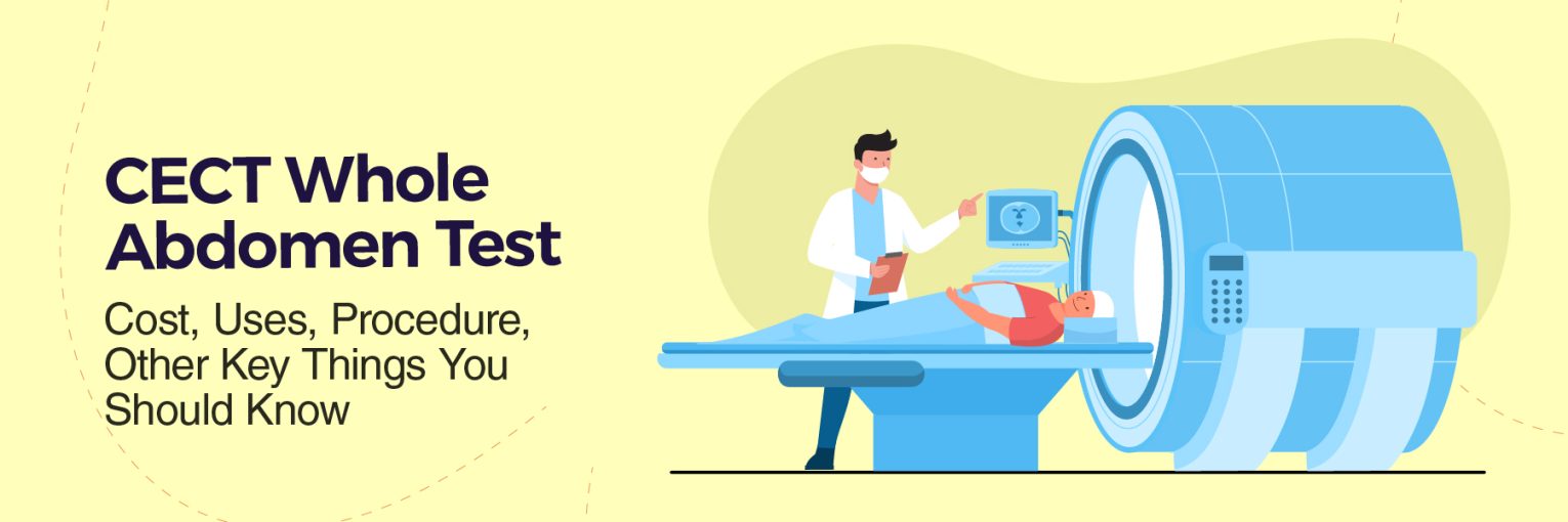
What Happens During the Test
Knowing what will happen inside the scanner can ease anxiety. The receptionist usually prints a sticker noting your CECT abdomen test time and attaches it to the request form.
Inside the suite, a radiographer explains breathing cues. Earplugs are unnecessary because modern scanners are quieter than older models. A small IV catheter is placed, and a saline flush confirms flow. The gantry’s lighted markings help align you symmetrically on the carbon-fibre table.
Typical Timeline
| Minute | Event |
| 0 | Positioning and scout image |
| 2 | Non-contrast pass |
| 4 | Contrast injection |
| 5 | Arterial acquisition |
| 7 | Portal venous acquisition |
| 12 | Delayed phase |
| 14 | Cannula removal, dressing |
| 15 | Preliminary technologist check |
An abdominal CECT may add an excretory phase at ten minutes if ureteric stones are suspected. Allow a small buffer around the scheduled CECT abdomen test time because emergency trauma cases occasionally receive priority.
- Throughout the scan, a two-way intercom keeps you in contact with staff. Lighting remains on, and you can see outside the ring, so claustrophobia is rare. You will hear a gentle whirring as the X-ray tube rotates; some people liken it to a washing machine’s spin cycle.
- Breath-hold commands last fewer than eight seconds. A mild metallic taste or warmth in the pelvis may occur as contrast circulates, both disappearing within a minute.
- Once off the table, you can dress immediately. Unless told otherwise, you are free to eat, drink and drive home, making the total clinic visit roughly thirty minutes.
Benefits and Risks
A single CECT scan of the abdomen can diagnose a ruptured spleen, hidden abscess or obscure tumour within minutes, and that speed can be lifesaving.
Major benefits
- Speed: Full acquisition in under ten seconds per phase
- Coverage: Organs, vessels, bones and lymph nodes in one study
- Availability: 24/7 in most tertiary hospitals
- Objectivity: Digital data allows precise tracking over time
Potential risks
- Radiation: 8–10 mSv, equal to three years of natural background exposure
- Contrast nephropathy: Risk rises when eGFR <30 mL min⁻¹; hydration mitigates
- Allergy: Mild hives in 3%, severe anaphylaxis in 0.02%
- Incidentalomas: Harmless nodules may prompt extra tests and worry
Putting numbers in context helps. For a 40-year-old, radiation-induced cancer risk is estimated at 1 in 2,000, whereas mortality from missed appendiceal rupture is closer to 1 in 10.
Technological advances continue to trim risk. Tube-current modulation, iterative reconstruction and organ-based shielding cut dose by up to 40% without compromising detail. Low-osmolar non-ionic contrast halves the chance of adverse reactions compared with older agents.
Always discuss kidney function, allergy history and pregnancy status with the radiologist. Together, you can decide whether the insight outweighs the hazard. In many urgent conditions, the answer is a confident yes, especially when alternative tests cannot deliver the same immediacy or scope. Refusal also carries risk; losing critical surgical minutes may prove more harmful than the modest radiation involved.
FAQs
Q1. What is a CECT abdomen scan used for?
Common CECT abdomen uses include diagnosing appendicitis, locating internal bleeding, staging cancers and checking for kidney stones.
Q2. Is fasting required before a CECT abdomen scan?
The CECT abdomen procedure typically relies on an empty stomach so that unexpected vomiting does not complicate contrast injection.
Q3. How much does a CECT abdomen scan cost in India?
The CECT test can cost anywhere from ₹4,500 in a district hospital to ₹12,000 in a premium urban centre.
Q4. Are there any side effects of a CECT abdomen scan?
Minor hives or a warm flush occur in about three per cent of people undergoing a CECT scan of abdomen, and serious reactions are extremely rare.
Q5. What is the difference between a CECT and a normal CT scan?
Both scans use X-rays, but CECT employs iodine contrast to highlight blood vessels and soft-tissue details that a plain CT might miss.