

Foot injuries are incredibly common, whether caused by sports, accidental falls, or overuse. In such cases, a detailed and accurate diagnosis is crucial to ensure proper treatment. One of the most commonly used imaging techniques for this purpose is the X-ray, especially in the Anteroposterior (AP) and Lateral views. These imaging methods provide clear and comprehensive visuals of the foot’s bones and joints. This article will help you understand what a Foot X-ray (AP & Lateral View) is, why it’s performed, and how it aids in detecting various foot conditions.
What is Foot X-ray (AP & Lateral View)?
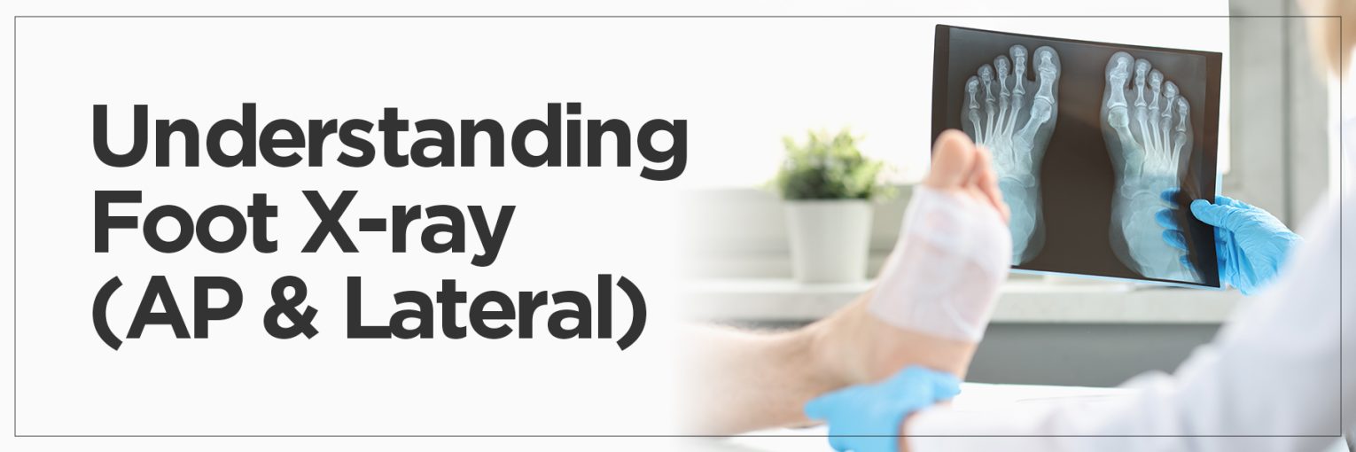
A Foot X-ray (AP & Lateral View) is a simple and painless imaging procedure that helps doctors see the structure of your foot from different angles. “AP” stands for Anteroposterior, meaning the X-ray beam passes from the front to the back of the foot, while “Lateral” means the image is taken from the side. Together, these two perspectives provide a complete view of the bones, joints, and soft tissues.
The X-ray foot AP lat helps identify problems with just one angle that might not be visible. For instance, if you’ve been experiencing pain, swelling, or can’t put weight on your foot, your doctor might recommend a foot X-ray to check for fractures, dislocations, or arthritis. A normal X-ray of foot will show aligned bones with no visible fractures or abnormalities. But if there’s any deviation from this, further treatment or investigation might be needed.
Purpose of Foot X-ray (AP & Lateral Views)
Doctors recommend Foot X-rays in AP and Lateral views for various reasons. One of the most common purposes is to identify fractures. Whether it’s a small stress fracture or a major break, an X-ray foot ap lat gives clear insight into the exact location and severity of the injury.
It is also essential in detecting xrays of broken foot to assess if bones have moved out of place or if surgical intervention is necessary.
Besides trauma, these views can help diagnose infections, arthritis, tumors, or abnormalities in bone growth. A regular foot X-ray might be part of a routine check-up for patients with chronic conditions like diabetes or osteoporosis.
The foot lateral X-ray is particularly useful in evaluating the arch and heel alignment, which is crucial in diagnosing flat feet or heel spurs. AP and Lateral foot X-rays are essential tools in orthopedic and podiatric care. They provide the images necessary for accurate diagnoses and effective treatment plans.
Procedure: How is a Foot X-ray Performed?
Getting a Foot X-ray is a quick, straightforward, and painless procedure. When you arrive at the radiology center or hospital, a technician will guide you to a special room where the imaging is done. First, you’ll be asked to remove your shoes and any jewelry or objects that could interfere with the image quality. For the X-ray foot AP lat view, you may be seated or standing, depending on the area to be examined and your ability to bear weight.
In the X-ray foot ankle AP lat procedure, the technician positions your foot flat for the AP (Anteroposterior) image, where the X-ray beam passes from front to back. Then, they will rotate your foot sideways for the lateral view to capture a side image. Usually, two to three images are taken to ensure a complete evaluation. The process takes only a few minutes, and you can resume normal activities immediately.
Diagnoses Made with Foot X-rays
Foot X-rays play a crucial role in diagnosing a variety of medical conditions. One of the most common reasons for performing a foot lateral X-ray is to detect minor stress fractures and more serious breaks. Additionally, doctors can identify dislocations, bone infections, and changes in bone structure that might indicate arthritis.
Other abnormalities, such as bone spurs, which are extra bone growths often linked to conditions like plantar fasciitis or arthritis, can also be spotted using different foot X-ray views. In children and adolescents, foot X-rays can help track bone growth and detect congenital deformities or alignment issues early on.
These images confirm a diagnosis and guide the treatment approach—whether it’s rest and immobilization, physical therapy, or surgery. By comparing current and previous X-rays, doctors can also assess how well an injury is healing.
Foot X-ray: Risks and Considerations
Foot X-rays are generally very safe, but there are a few risks and considerations to consider. One of the main concerns with any X-ray is exposure to a small amount of ionizing radiation. Although minimal, repeated exposure over time should be avoided unless necessary. During a foot AP lat view X-ray, precautions such as protective lead shields may be used to limit radiation to other body parts.
Pregnant women should inform their doctor or technician before undergoing any radiography, as fetal exposure to radiation should be minimized or avoided. In such cases, alternative imaging methods or delayed procedures may be considered, depending on urgency.
It’s also important to remain still during the scan to ensure a clear image. Blurry or unclear images may require a repeat, leading to additional exposure. For most individuals, the benefits of accurate diagnosis far outweigh the minimal risks associated with a standard foot X-ray.
FAQs
What can an X-ray of your foot show?
A foot X-ray can reveal various bone-related issues, including fractures, dislocations, arthritis, bone spurs, and congenital deformities. It helps doctors examine the alignment and integrity of the bones and joints in the foot. It’s a crucial first step in diagnosing injuries or abnormalities caused by trauma, overuse, or chronic conditions.
Can X-rays show foot pain?
X-rays don’t directly show pain, but they can reveal the cause behind the discomfort. For example, if foot pain is due to a fracture, bone spur, or arthritic changes, the X-ray will highlight these abnormalities. However, if the source of the pain is related to soft tissue injuries like ligament tears or plantar fasciitis, these won’t show up clearly on a standard X-ray.
Can an X-ray show infection in foot?
Yes, in some cases. While X-rays can detect signs of infection, such as bone erosion, joint space narrowing, or bone destruction, early-stage infections might not be visible. If an infection is suspected but not confirmed through X-ray, additional imaging like an MRI may be recommended for a more detailed assessment.
Why does my foot hurt, but the X-ray is normal?
Sometimes, a foot can hurt even when the X-ray appears normal. This may happen in soft tissue injuries, nerve problems, early stress fractures, or conditions like plantar fasciitis. These are not always visible on X-rays. Further diagnostic imaging, like ultrasound or MRI, may be necessary.
What scan is best for foot pain?
While X-rays are ideal for bone issues, MRI is the best scan for foot pain related to soft tissues, nerves, or undetected stress fractures. Ultrasound is also useful for certain ligament or tendon issues.