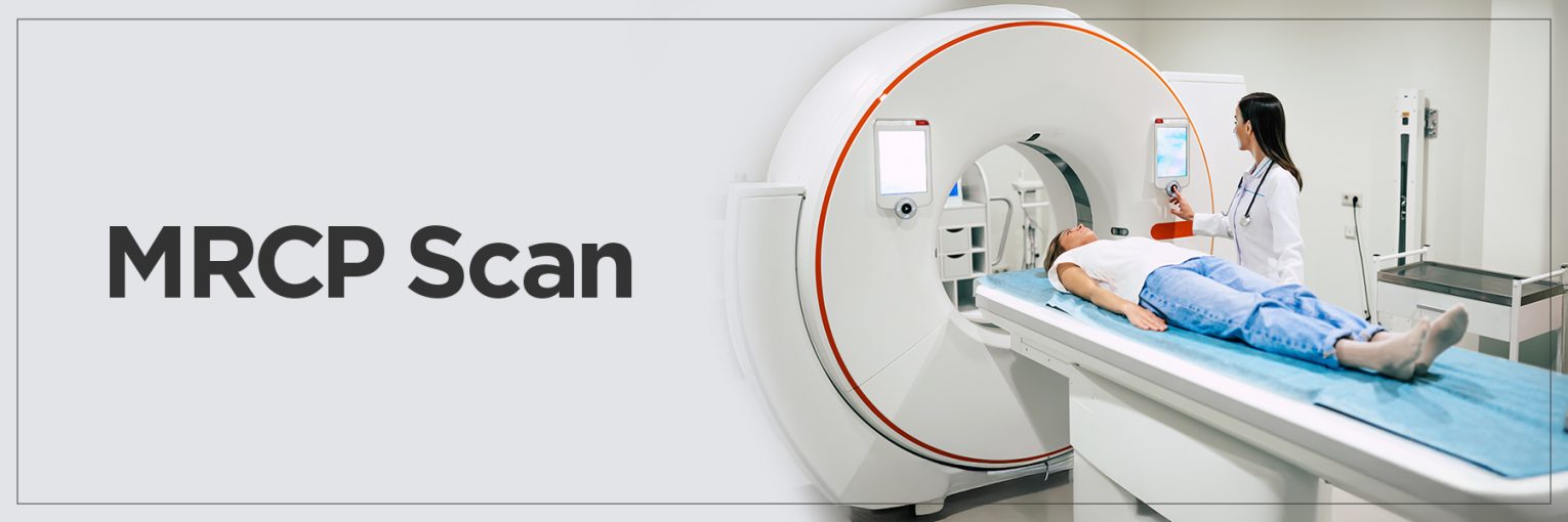
Medical imaging has transformed the detection and diagnosis of any health problem inside the body. An example of such a technique is the MRCP scan, which is a specific form of MRI that produces high-quality images of the bile ducts, gallbladder, pancreas, and liver.
This noninvasive scan, which requires a 1.5T MRI machine, detects gallstones, blockages, inflammation, and more, eliminating the need for surgery and endoscopy.
In most cases, it is a safe, painless procedure that does not require the use of contrast dye. This information guide describes an MRCP scan, the reasons it is performed, how it works, and what patients should expect during the test.
What is an MRCP Scan?
An MRCP scan, also known as magnetic resonance cholangiopancreatography, is a noninvasive imaging procedure designed to view the bile ducts, pancreatic duct, liver, and gallbladder.
Most of the time, the MRCP MRI scan is a safer and more pleasant substitute for conventional diagnostic techniques such ERCP (Endoscopic Retrograde Cholangiopancreatography) since it does not involve surgery or contrast agents.
Since this scan clearly depicts blockages or narrowing of the bile or pancreatic ducts, doctors frequently advise it to patients with unexplained abdominal discomfort or jaundice. Owing to its accuracy and non-invasiveness, the MRCP scan has evolved into a critical diagnostic instrument in detecting early-stage conditions without pain or protracted recovery periods.
Purpose of an MRCP Scan
The MRCP scan is mostly employed to identify issues with the liver, pancreas, and bile ducts.
- One of the most often used MRCP indications is to look into the source of jaundice, abdominal pain, or abnormal liver function tests.
- It can effectively find gallstones, inflammation, stricture, or tumors within the bile or pancreatic ducts, as well as bile duct obstructions.
- Other MRCP uses include inspecting for congenital defects of the ducts, assessing chronic pancreatitis, or monitoring patients with a past history of gallbladder problems.
- It is particularly advantageous for those who might not withstand invasive surgery. It is usually chosen for initial evaluation as the scan is non-invasive and does not typically call for contrast agents.
MRCP scans give physicians a safe and dependable view of the internal duct systems, aiding in their diagnosis of significant diseases without surgical hazards or problems.
Procedure: How is an MRCP Scan Performed?
MRCP testing is straightforward, non-invasive, and typically lasts between 30 to 60 minutes.
- Before the scan, patients may be asked to fast for a few hours to ensure the gallbladder and ducts are clearly visible.
- Upon arrival, they will change into a gown and remove any metal objects because they cause issues with the magnetic field.
- For the scan itself, the patient lies down on a table that slides into the MRI machine.
- During the MRCP imaging technique, the machine uses powerful radio waves and magnets to capture detailed images of the biliary and pancreatic ducts.
- Patients must remain still and may be asked to hold their breath briefly during certain image captures to avoid blurring.
A common question is: “Why do I need an MRCP scan?” If you’re experiencing unexplained digestive issues, pain, or abnormal lab results, your doctor may order this test to get a clearer view of potential internal problems.
Diagnosis Made with MRCP Scans
The MRCP MRI scan is instrumental in diagnosing a variety of conditions that affect the biliary and pancreatic systems.
- Among the most serious diagnoses it can assist with is cholangiocarcinoma, a rare but aggressive cancer of the bile ducts. By providing high-resolution images, MRCP helps identify tumors, strictures, or irregularities that may suggest malignancy early on.
- Another common condition detected through MRCP testing is pancreatitis, both acute and chronic forms. MRCP helps to learn about swelling, inflammation, and structural damage in the pancreas. It gives insights into the seriousness and cause of the condition.
- The scan is also invaluable in detecting choledocholithiasis. It refers to gallstones that have got into the bile duct. They can cause infection, pain, or blockages if not promptly treated.
- MRCP can reveal congenital anomalies of the biliary tract, post-surgical complications, and signs of autoimmune diseases affecting the liver or pancreas.
Thanks to its non-invasive nature and accuracy, MRCP is a preferred first-line diagnostic approach for physicians evaluating patients with complex abdominal symptoms or uncertain diagnoses.
MRCP Scan – Risks and Considerations
Though the MRCP MRI scan is generally considered safe and non-invasive, there are a few risks and considerations patients should be aware of.
- The strong magnetic fields used in the scan mean it’s unsuitable for individuals with certain implants, such as pacemakers, cochlear implants, or metal fragments in the body. Always inform the technician if you have any such devices before scheduling your MRCP scan.
- In rare cases where a contrast agent is needed (though MRCP typically does not use one), patients with allergies to gadolinium-based agents may experience side effects, including nausea, dizziness, or skin reactions.
- Those with kidney issues should be assessed carefully before receiving contrast, due to the potential risk of nephrogenic systemic fibrosis.
- While MRCP testing is not associated with radiation exposure—unlike CT scans or X-rays—it can be uncomfortable for patients who are claustrophobic, as it requires lying still in an enclosed MRI tube.
Some centers offer open MRI machines or mild sedation to help manage this. Overall, the benefits of MRCP far outweigh the risks, especially when used for the timely and accurate diagnosis of potentially serious conditions.
MRCP vs. ERCP: Comparing Diagnostic Tools
When it comes to diagnosing issues of the bile and pancreatic ducts, both MRCP testing and ERCP (Endoscopic Retrograde Cholangiopancreatography) are effective but serve different purposes.
- MRCP uses magnetic resonance technology to create detailed pictures, ideal for initial diagnosis and evaluation. On the other hand, ERCP test is an invasive procedure that combines endoscopy and X-ray imaging to not only diagnose but also treat problems within the ducts.
- MRCP is typically the first choice when doctors want to visualize duct abnormalities without intervention. It avoids the risks associated with sedation, infection, or pancreatitis—common complications with ERCP.
- While MRCP can show stones, strictures, or tumors, it cannot remove them or take tissue biopsies. That’s where ERCP becomes valuable. If a therapeutic intervention is needed—such as stent placement, stone removal, or biopsy—ERCP is the preferred tool.
- MRCP offers a safe, detailed, and non-invasive way to evaluate internal organs, making it ideal for diagnosis. ERCP, while more invasive, becomes essential when diagnosis and treatment need to happen simultaneously.
Choosing between the two depends on the clinical goal—observation or intervention.
FAQs
1. What is the goal of MRCP?
The primary goal of MRCP (Magnetic Resonance Cholangiopancreatography) is to provide clear, detailed images of the bile ducts, pancreatic duct, liver, and gallbladder without the need for surgery or invasive procedures.
It helps doctors diagnose a variety of conditions, including blockages, inflammation, gallstones, strictures, and tumors. MRCP is particularly useful in evaluating unexplained abdominal pain, jaundice, or abnormal liver enzyme levels.
2. What is the difference between an MRI and an MRCP scan?
MRI (Magnetic Resonance Imaging) is a broad imaging technique used to visualize various internal organs and tissues throughout the body using magnetic fields and radio waves. MRCP is a specialized form of MRI that focuses specifically on the hepatobiliary and pancreatic systems.
While a standard MRI may assess the brain, spine, joints, or soft tissues, MRCP is tailored to capture sharp images of the bile ducts, pancreas, and surrounding organs.
3. Does MRCP show gallstones?
Yes, MRCP is highly effective at detecting gallstones, particularly those located in the common bile duct—a condition known as choledocholithiasis.
While ultrasound is often used initially to detect gallstones within the gallbladder, MRCP provides a more comprehensive view of the entire biliary tree, making it particularly useful for identifying stones that have migrated beyond the gallbladder.
4. Can MRCP detect a tumour?
Yes, MRCP can detect tumors, especially those affecting the bile ducts, pancreas, or liver. It is particularly helpful in identifying cholangiocarcinoma (bile duct cancer), pancreatic cancer, and masses that cause obstruction or ductal dilation.
MRCP may not always provide definitive confirmation of a tumor’s nature, but it can reveal abnormal growths or irregularities that warrant further investigation, often leading to a biopsy or follow-up imaging for a precise diagnosis.
5. What is the scope after MRCP?
After an MRCP scan, your doctor will review the results to determine the next steps. If abnormalities are found, you may be referred to a specialist such as a gastroenterologist or surgeon. Depending on the findings, further diagnostic tests like ERCP, CT scan, or biopsy may be recommended. In some cases, therapeutic procedures or surgery may follow.


