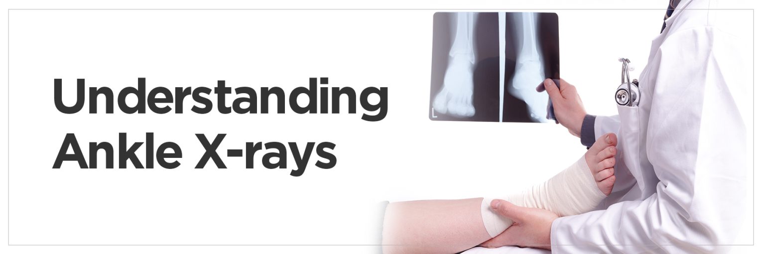
When an ankle injury happens — whether from a fall, twist or sports-related mishap — the first step in diagnosis is often an X-ray. An X-Ray Ankle AP & Lateral View is a basic imaging test that allows doctors to view inside the ankle joint without having to make even one incision. It takes little time, poses no harm to the patient, and is particularly good at detecting fractures, dislocated bones and joint abnormalities.
The two major views of the ankle that allow us to see the bones and other structures in this region are the AP (Anteroposterior) and Lateral views. Understanding this is critical if you suffer from persistent ankle pain, swelling or trauma. Here’s what you need to know about this X-ray, why it’s used and how it’s done.
What is an X-Ray Ankle AP & Lateral View?
An X-Ray Ankle AP & Lateral View is a type of two-angle imaging examination that aims to understand the structure of the ankle. An ankle AP X-Ray (Anteroposterior) is a front-to-back view of the leg capturing two long bones (the tibia and fibula) and the talus in a straight line. This perspective gives a better idea of the position of the ankle joints and the spaces between the bones.
The ankle lateral view, in contrast, is taken laterally, allowing depth and more information on the proximity of the bones. When combined, these images — known collectively as the ankle ap lateral — provide a balanced assessment.
They play a vital role in identifying fractures, dislocations, and degeneration, providing physicians with an idea of the next steps in treatment. If a soft tissue injury is suspected, more detailed scans, such as MRIs, are typically done after these X-rays.
Why is an Ankle X-Ray Needed?
An X-ray of the ankle becomes necessary when there’s pain, swelling, or visible deformity following an injury. It’s primarily used to diagnose fractures, which are common after twists or direct impacts. It helps determine the type and location of the fracture—be it in the tibia, fibula, or talus—and how severe it is. X-rays can also reveal ankle ligament injuries indirectly when bones appear displaced or misaligned.
However, for detailed ligament analysis, doctors may follow up with an ankle ligament MRI, especially if no fractures are seen but instability persists.
In addition to trauma, X-rays are useful in evaluating long-standing pain or swelling due to arthritis, which causes joint space narrowing and bone spurs. They can detect signs of osteomyelitis, a serious bone infection, and tumours or cysts in rare cases.
When swelling or pain continues without a clear cause, an ankle injury MRI might be considered next. But, the AP and Lateral X-ray views remain the cornerstone of initial ankle diagnostics, helping to quickly identify or rule out serious conditions and guide the next steps in treatment.
How is an X-Ray Ankle AP & Lateral View Done?
The X-ray procedure for the ankle AP and lateral view is simple, fast, and painless. It begins with the patient being asked to remove any metallic items that could interfere with image clarity. The patient is typically seated or lying on a table in the radiology room, and a lead apron may be used to shield the body from unnecessary radiation.
For the Ankle AP X-ray view, the foot is placed flat with the toes pointing upward, allowing the X-ray beam to pass from the front to the back of the ankle. The technician ensures the correct angle to capture the bones’ frontal alignment. In the ankle lateral view, the patient turns the leg outward, or lies on the side, so the beam can pass from the inner to the outer side of the ankle.
- These ankle X-ray views are critical in visualising the joint from different angles, ensuring no abnormalities are missed.
- The X-ray machine emits a controlled burst of radiation, which passes through the ankle and is captured on a digital detector or film on the opposite side. The entire procedure takes about 10–15 minutes. Once the images are captured, a radiologist interprets them and sends a report to the referring physician.
- This diagnostic test is highly valuable for confirming injuries, planning surgeries, or monitoring recovery. It’s a non-invasive and widely available imaging option with minimal risks, making it a preferred choice in most clinical settings.
Patient Preparation for Ankle X-Ray
Proper Ankle X-Ray Preparation ensures clear imaging and patient safety. Before the scan, the patient is asked to remove any clothing, shoes, or accessories covering the ankle. Loose, comfortable clothing is usually preferred, and a gown might be provided. Any metal objects—including zippers, jewelry, or buttons near the ankle—should be removed as they can obstruct the image quality.
Patients should inform the technician if they are pregnant or suspect pregnancy, as precautions may be taken to reduce exposure. While many wonder, “is X-Ray safe?” the answer is yes—the radiation used is minimal and controlled. In fact, a typical ankle AP x-ray delivers a radiation dose far below harmful thresholds.
Lead aprons or shields are often used to protect other parts of the body during the scan. Following these preparation steps allows for a smooth and accurate imaging session, helping the technician capture the best possible views.
What Happens During the X-Ray?
The process of X-ray for an ankle AP & Lateral view is simple and generally painless, making it one of the most common diagnostic tools for orthopedic concerns. Once the patient arrives at the imaging centre, they are escorted to the X-ray room and asked to remove shoes, socks, and any jewellery or metal objects near the ankle.
A lead apron may be provided to shield parts of the body from being scanned, especially in cases where radiation exposure must be minimised.
For the AP (Anteroposterior) view, the patient is asked to place their foot flat on the table with the toes pointing upwards. This angle helps capture the bones of the ankle in a straight line from front to back. Then, for the Lateral view, the foot is repositioned sideways—either while the patient lies on their side or turns the leg outward—so the X-ray beam can pass from the inner side to the outer side of the ankle.
The technician ensures the correct position, steps behind a protective screen, and activates the machine. Each exposure takes only a fraction of a second. Patients are asked to stay completely still to prevent any image blurring. If necessary, additional angles may be taken.
Throughout the procedure, X-ray safety precaution guidelines are strictly followed. The radiation used is minimal, but shielding and equipment calibration help ensure patient safety.
The whole process takes approximately 10–15 minutes, and most people find it quick, efficient, and completely tolerable, even when in mild pain from an injury.
FAQs on Ankle X-Ray
Q1. What is an X-Ray Ankle AP & Lateral View?
An X-Ray Ankle AP & Lateral View is a diagnostic imaging test that captures two different angles of the ankle: AP (Anteroposterior) and Lateral. The AP view is taken from the front, while the Lateral view is taken from the side. These perspectives help doctors detect fractures, dislocations, or joint irregularities by offering a comprehensive visual of the bones and joint space.
Q2. How long does an ankle X-ray take?
The entire X-ray procedure usually takes 10 to 15 minutes. This includes positioning, capturing the AP and Lateral views, and sometimes repeating the images if necessary. The imaging itself lasts only a few seconds, but careful positioning ensures accuracy.
Q3. Are there any risks associated with an ankle X-ray?
X-rays use low levels of ionising radiation, which is generally considered safe for most people. Risks are minimal, especially when appropriate precautions like lead aprons are used. However, pregnant women should inform their healthcare provider in advance so alternative methods or extra shielding can be considered.
Q4. What can an ankle X-ray detect?
An ankle X-ray can detect fractures, dislocations, bone infections (osteomyelitis), arthritis, bone spurs, and even signs of tumours. It is also used to monitor healing after surgery or injury. While it doesn’t show soft tissues in detail, it may suggest indirect signs of ligament damage, prompting further tests like an MRI if needed.
Q4. How long does an ankle X-ray take?
The entire X-ray procedure usually takes 10 to 15 minutes. This includes positioning, capturing the AP and Lateral views, and sometimes repeating the images if necessary. The imaging itself lasts only a few seconds, but careful positioning ensures accuracy.
Q5. Are there any risks associated with an ankle X-ray?
X-rays use low levels of ionising radiation, which is generally considered safe for most people. Risks are minimal, especially when appropriate precautions like lead aprons are used. However, pregnant women should inform their healthcare provider in advance so alternative methods or extra shielding can be considered.
Q6. What can an ankle X-ray detect?
An ankle X-ray can detect fractures, dislocations, bone infections (osteomyelitis), arthritis, bone spurs, and even signs of tumours. It is also used to monitor healing after surgery or injury. While it doesn’t show soft tissues in detail, it may suggest indirect signs of ligament damage, prompting further tests like an MRI if needed.


