

Abdominal Magnetic Resonance Imaging (MRI) is a non-invasive diagnostic tool that generates detailed images of the abdominal organs and tissues using a strong magnetic field, radio waves, and computer technology. An MRI Abdomen scan is especially helpful when doctors require a precise picture of soft tissues that alternative methods cannot assess with conventional X-rays or CT images.
Offering a high degree of accuracy without subjecting patients to ionising radiation, this test is essential for detecting, diagnosing, and tracking many different abdominal problems. An abdomen MRI scan is especially helpful for patients with undiagnosed stomach symptoms or for monitoring the development of identified medical diseases.
What is an MRI Abdomen Scan?
Designed specifically, an MRI Abdomen scan aids doctors in examining the structures and organs inside the abdominal cavity, including the liver, kidneys, pancreas, spleen, and intestines. An abdomen MRI scan generates high-resolution, cross-sectional images using strong magnetic fields and radio waves rather than X-rays or CT scans; therefore, it is particularly well-suited to detect minute abnormalities.
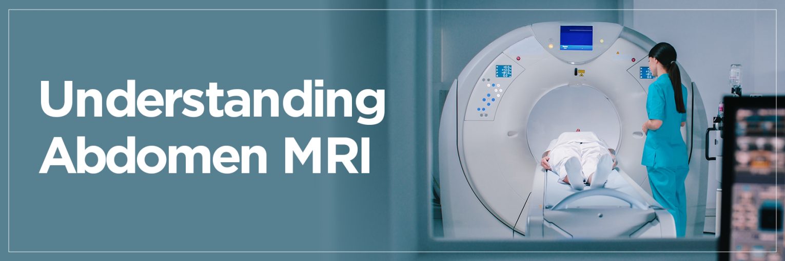
During an abdomen MRI scan, the patient rests on a table that glides into a big tube-shaped machine. Depending on the complexity of the condition being examined, the scan may last 30 to 60 minutes.
Some doctors recommend an MRI whole abdomen to obtain a complete picture of the area. This aids in the detection or ruling out of systemic disorders, evaluation of organ capacity, and highly accurate guidance of treatment plans.
Why is an MRI Abdomen Test Done?
An abdomen MRI scan is usually recommended when a patient’s symptoms cannot be totally clarified by either a physical examination or simple imaging tests. This covers persistent or unexplained stomach pain, bloating, or digestive problems. MRI scans are most often used to diagnose liver disorders such as liver lesions, cirrhosis, or fatty liver. They also greatly help assess kidney diseases, including cysts, stones, and tumours.
Diagnosing pancreatic diseases, including pancreatic cancer or inflammation (pancreatitis), is yet another critical use. Doctors also use the MRI scan stomach to evaluate stomach wall abnormalities, find ulcers, or look for gastric cancer indicators.
A triple-phase MRI abdomen may be done for more thorough and accurate liver imaging, particularly when assessing lesions or tumours. This helps doctors more accurately distinguish between benign and malignant tumours by imaging three distinct phases of contrast dye circulation – arterial, venous, and delayed.
MRI of the abdomen is a crucial tool that can expose a wide range of medical disorders, therefore guaranteeing prompt and adequate medical treatment.
MRI Abdomen: Step-by-Step Procedure
The normal abdomen MRI usually starts with a comprehensive process explanation by a radiologist or technician.
- The patients are commonly instructed to steer clear of food and drinks for several hours prior to the test.
- Since the magnetic field might affect devices, it is also vital to notify the technician of any implants, pacemakers, or metal items within the body.
- The patient lies down on a motorised table and changes into a hospital gown during the MRI abdomen operations.
- A coil (a specific device used to enhance image quality) could be placed over the abdominal region.
- Should contrast dye, usually gadolinium, be needed, it is injected via an intravenous (IV) line in the arm. This dye helps to highlight organs, tissues, and blood arteries, therefore improving the spotting of anomalies.
- Once the patient is ready, the table moves into the MRI scanner. The loud tapping or thumping noises the machine generates are completely normal.
- Patients frequently receive headphones or earplugs to help them block out the noise. However, to guarantee the images are clear and correct, one must remain motionless throughout the scan.
- If contrast dye is used after the scan, the patient might be observed for a brief period to eliminate allergic responses.
- Usually, patients can start doing their regular things right away. A radiologist reviews the scan results and sends them to the referring physician, who will then go over the conclusions.
Understanding the MRI abdomen procedure helps patients to gain confidence and lower their worry as they know what to anticipate during the process.
How to Prepare for an MRI Abdomen Scan?
Proper preparation is essential to ensure the accuracy and safety of your abdomen MRI scan.
- Patients are typically advised to avoid eating or drinking for 4 to 6 hours prior to the scan, particularly if contrast dye will be used. This fasting helps reduce motion in the digestive tract, allowing for clearer imaging results.
- Additionally, any metal objects such as jewelry, piercings, or hearing aids must be removed, as they can interfere with the magnetic field used in the scan.
- During MRI preparation abdomen, patients should inform their radiologist if they have any implants, such as pacemakers or metal clips from previous surgeries, as these could pose risks. For those requiring contrast dye, it’s important to alert the medical team about any allergies, kidney problems, or past reactions to contrast agents.
These precautions ensure a smooth and safe MRI experience while enhancing the diagnostic clarity of the images.
What Happens During the MRI Scan?
When it’s time for the MRI scan of the stomach, you’ll be guided into the scanning room and asked to lie flat on a table. A coil may be placed around your abdomen to improve the imaging quality. If your doctor has requested an MRI upper abdomen with contrast, you’ll receive an intravenous injection of gadolinium, a special dye that highlights blood flow and organ detail.
Once you’re settled, the table slides into the abdominal MRI machine, which resembles a large tube. The machine will make rhythmic tapping or thudding sounds during the scan, so earplugs or headphones are often provided for comfort. It’s crucial to remain very still, as movement can blur the images.
The scan usually lasts between 30 to 60 minutes. Though the environment may feel confined, the process is painless. If you experience anxiety or claustrophobia, sedation may be offered. Once the scan is complete, you can resume your daily routine almost immediately.
Benefits of an MRI Abdomen Scan
- One of the most significant benefits of undergoing an MRI upper abdomen with contrast is the ability to obtain detailed, high-resolution images without using ionising radiation. This makes it safer, especially for repeated imaging or for vulnerable groups like pregnant women and children.
- Unlike X-rays and CT scans, which primarily detect bones and dense structures, an MRI provides sharp views of soft tissues, organs, and blood vessels. This makes it particularly effective in diagnosing liver diseases, pancreatic tumours, kidney abnormalities, and other internal issues.
- The abdominal MRI machine is designed to create cross-sectional views, allowing doctors to see different layers and perspectives of the abdominal cavity. This enhances diagnostic accuracy and enables early detection of complex conditions.
- Moreover, an MRI abdomen scan is non-invasive and painless, eliminating the need for exploratory surgery in many cases. It can be customised with or without contrast dye, depending on the diagnostic need. With its exceptional clarity and safety profile, MRI has become the preferred imaging method for detailed abdominal analysis.
MRI Abdomen vs CT Scan vs Ultrasound
When comparing imaging methods, each has its own strengths.
- An MRI Abdomen scan provides superior soft tissue contrast, making it ideal for evaluating organs such as the liver, kidneys, pancreas, and intestines. It’s particularly beneficial when the diagnosis demands highly detailed images without exposure to radiation, such as in cancer detection or vascular mapping with contrast.
- CT scans, on the other hand, are faster and more widely available. They are often used in emergency situations, such as trauma, where quick imaging is critical. CT scans use ionising radiation, which may not be suitable for all patients, particularly during pregnancy or for repeated use.
- Ultrasound is a low-cost, portable, and radiation-free method best suited for real-time imaging of moving structures, such as gallbladder function or fetal development. However, due to its lower image clarity, it is limited to deeper organs or obese patients.
If you’re searching for an “ultrasound near me,” it’s likely because it’s often the first step in abdominal imaging due to its accessibility. But for comprehensive internal visualisation, especially when symptoms are unclear or persist, doctors may recommend an MRI Abdomen scan to get a clearer, more accurate picture.
How to Book an MRI Abdomen Scan?
Booking an MRI Abdomen scan is now easier than ever. You can book it online through trusted diagnostic centres by simply selecting your city, preferred date, and test type. One of the top recommendations is Vijaya Diagnostic Centre, known for 40+ years of excellence in healthcare diagnostics.
With over 160 diagnostic centres and a team of 200+ professionals, including radiologists, pathologists, and microbiologists, Vijaya Diagnostic Centre serves more than 50 million customers and 250+ corporate clients.
Vijaya Diagnostics is trusted by patients of every age for its state-of-the-art facilities and best-in-class customer service. When comparing options like “MRI Abdomen” or “ultrasound near me,” Vijaya stands out for quality and trust.
Before booking your abdomen MRI scan, check if your health insurance covers the test. Most major providers do. Online booking platforms often allow you to upload prescriptions, choose add-on services, and make secure payments in a few easy steps.
FAQs on MRI Abdomen Scan
1. What is an MRI 1.5 Whole Abdomen scan?
An MRI 1.5 Whole Abdomen scan uses a 1.5 Tesla strength MRI machine to produce detailed cross-sectional images of all major abdominal organs. It is commonly recommended to evaluate the liver, kidneys, pancreas, spleen, adrenal glands, and gastrointestinal tract. The scan offers precise visuals without radiation, making it ideal for diagnosing complex or chronic abdominal conditions.
2. How long does an MRI 1.5 Whole Abdomen scan take?
An MRI 1.5 Whole Abdomen scan typically takes about 30 to 60 minutes, depending on the need for contrast dye or additional sequences. The process includes patient setup, the actual scanning, and quality checks. If contrast is administered, a short wait time is added to ensure proper distribution throughout the abdominal area for clearer imaging.
3. Is an MRI Whole Abdomen scan safe?
Yes, an MRI Whole Abdomen scan is considered very safe. It uses strong magnetic fields and radio waves, not radiation, making it preferable over X-rays or CT scans, especially for children and pregnant women. The scan is non-invasive, painless, and suitable for regular monitoring of chronic conditions without long-term health risks when guidelines are properly followed.
4. Are there any side effects of an MRI Whole Abdomen scan?
Side effects from an MRI Whole Abdomen scan are rare. Some patients might feel discomfort from lying still or experience mild reactions to the contrast dye, such as nausea or a metallic taste. Serious allergic reactions are extremely uncommon. People with metal implants should consult their doctor beforehand, as the magnetic field can interfere with such devices.
5. What is the difference between an MRI and a CT scan for the abdomen?
An MRI provides superior soft tissue contrast and doesn’t use ionising radiation, making it ideal for liver, pancreas, and kidney evaluations. CT scans are faster and more accessible, and are often used in emergency situations like internal injuries. However, CT involves radiation and is less detailed for soft tissues compared to MRI, which offers higher resolution but takes longer.
MRI Scan Centre
To find MRI Scan Centre near me click here.