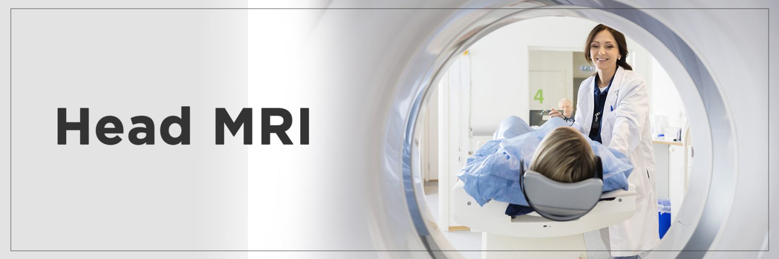
A head MRI is a potent diagnostic tool that gives doctors a sharp view of what’s going on inside your brain without a single cut. A Head MRI can generate thorough images to help identify the cause, whether you suffer from long-term headaches, dizziness, or seizures.
With the sophisticated technology, this non-intrusive scan aids in the identification of strokes, inflammation, tumors, and other problems. When you know what to expect, it can help you feel more ready and calm your agitation.
This guide will explain what a Head MRI is, how it works, when it is advised, and what your results could mean. We will explore this contemporary wonder of medical imaging.
What is a Head MRI?
Using powerful magnetic fields and radio signals, a head MRI creates very specific pictures of the brain and surrounding tissues. It is a medical imaging technique. Although X-rays or CT scans use ionizing radiation, an MRI does not, so it’s a better choice for frequent or in-depth imaging. A head MRI is mainly used to diagnose or track neurological disorders, evaluate brain injuries, or identify variations, including tumors, hemorrhages, or infections.
By enabling doctors to see the brain from many points, the MRI scanning procedure helps offer better diagnoses. It is especially good for spotting small changes in soft tissues that other imaging techniques might not clearly show. Patients can find solace in knowing the MRI scan meaning; it is not only about the pictures but about how they reveal opportunities for recovery and therapy.
How Does a Head MRI Work?
The theory behind a Head MRI is amazing. The equipment aligns hydrogen molecules in the body using a strong magnetic field. Once aligned, the MRI machine sends radiofrequency pulses that temporarily disturb this alignment. The atoms return to their starting positions and produce energy when the radio waves are turned off. Receivers grab this energy, and software programs transform it into pictures.
All of this enables the machine to tell apart several tissue types, which in turn assists physicians and radiologists distinguishing between normal and abnormal brain MRI findings. If you ever wanted to know how MRI works, it is all about magnetic physics and radio waves acting in tandem. Since it offers more contrast between healthy and sick tissue than other scans, an MRI is especially good for soft tissue imaging.
When is a Head MRI Recommended?
Doctors may recommend a Head MRI when you experience symptoms that suggest a neurological issue. These can include persistent headaches, unexplained dizziness, seizures, changes in vision, or cognitive difficulties. A head MRI is often used to evaluate these symptoms further and confirm a diagnosis.
For brain imaging, you might wonder which is better, MRI or CT scan for brain conditions. While CT scans are faster and excellent for detecting acute bleeding or skull fractures, MRI head scans offer far greater detail, especially for soft tissues like the brain. MRIs are particularly recommended when conditions such as multiple sclerosis, brain tumors, or developmental anomalies are suspected.
Ultimately, a doctor’s recommendation will depend on your symptoms, medical history, and the urgency of your condition. A head MRI is a valuable tool for getting a closer look at the brain when clarity is needed most.
What Does a Head MRI Show?
A head MRI can uncover a wide range of medical conditions that affect the brain.
- Some of the most common findings include brain tumors, stroke, aneurysms, multiple sclerosis, traumatic brain injuries, and infections like meningitis.
- It can also detect degenerative diseases such as Alzheimer’s and Parkinson’s, as well as congenital abnormalities in the brain’s structure.
- When asking what MRI can detect, it’s important to understand that this imaging tool is incredibly sensitive. It can identify even minute changes in brain tissue, blood vessels, and fluid levels.
- An MRI head scan can reveal swelling, bleeding, or structural defects that might not show up in other imaging tests.
This detailed view makes a head MRI an essential step in diagnosing serious conditions, monitoring the progress of ongoing brain disorders, or planning surgeries. With early detection, treatment can begin sooner, improving outcomes significantly.
How Do I Prepare for a Head MRI?
Preparation for an MRI procedure is simple but important. You’ll typically be asked to wear loose-fitting, comfortable clothing that doesn’t contain any metal—this includes zippers, buttons, or underwire bras. If your clothing isn’t suitable, a hospital gown may be provided. Removing all jewelry, watches, hairpins, eyeglasses, hearing aids, and any electronic items is crucial, as metal can interfere with the magnetic field and compromise image quality.
Depending on the purpose of MRI scan, your doctor may advise fasting for a few hours before the test, especially if a contrast agent is going to be injected. This contrast dye, usually gadolinium-based, helps enhance the images for a more accurate diagnosis. Your healthcare provider will tell you if any dietary restrictions apply.
If you’re undergoing an MRI head scan, it’s also wise to inform your doctor about any implants, pacemakers, or medical conditions like kidney problems or claustrophobia. Following these guidelines ensures a smooth experience and optimal image clarity. Here are some things to keep in mind –
| Aspect | Before the MRI | After the MRI |
| Clothing | Wear loose-fitting clothes without metal (no zippers, buttons, or underwire). | You can change back into your regular clothes. |
| Jewelry & Accessories | Remove all metal objects: jewelry, hairpins, eyeglasses, hearing aids, watches, etc. | Rewear your personal items once cleared by the technician. |
| Electronic Devices | Leave phones, credit cards, and electronic devices outside the scan room. | Retrieve and use your devices as normal after the test. |
| Medication | Continue regular meds unless told otherwise by your doctor. | Continue medication unless post-scan instructions say otherwise. |
| Medical History Disclosure | Inform technician of pacemakers, metal implants, allergies, or kidney conditions. | Watch for any delayed reaction (rare) if contrast was used. |
| Mental Preparation | Prepare for 30–60 minutes in a confined space; discuss anxiety or claustrophobia with the doctor. | You may feel slightly disoriented if sedatives were used—rest if needed. |
What Should I Expect During a Head MRI?
Knowing what to expect during a brain MRI can ease your nerves.
- You’ll be asked to lie down on a narrow table, which then slides into a large, tube-like machine.
- The MRI scanner comes with a powerful magnetic field, so you’ll hear loud knocking or thumping noises during the imaging—don’t worry; this is completely normal. Earplugs or headphones are typically provided to minimize the sound.
- Throughout the MRI process, you must remain perfectly still to ensure clear images.
- Depending on the facility, the scan might use a 1.5T MRI (Tesla), which is a standard strength that offers excellent image detail for diagnosing neurological issues.
- The procedure is painless, although some people may feel confined inside the scanner.
The scan is designed to capture both normal and abnormal brain MRI details, helping doctors assess everything from structure to signs of disease. A technician monitors you from another room and can communicate via intercom, offering reassurance and instructions throughout.
How Long Does a Head MRI Take?
If you’re wondering how long does an MRI take, the average Head MRI lasts between 20 to 30 minutes. The time depends on the specific images your doctor needs, whether a contrast agent is being used, and how many sequences are required. A standard scan without contrast is generally shorter, while a contrast-enhanced MRI may take longer due to the injection and additional imaging.
It’s important to arrive a little early for paperwork and pre-scan procedures. Once the head MRI begins, lying still is critical, as movement can blur the images and require re-scanning.
Most people tolerate the scan well, though some may experience mild MRI scan side effects like dizziness or a metallic taste if contrast is used. These effects are rare and usually short-lived. If you feel anxious or claustrophobic, let the staff know—they can offer support or even mild sedation if needed.
FAQs
1. Can MRI detect brain damage?
A. Yes, MRI is one of the most accurate tools for detecting brain damage. It can identify signs of trauma, bleeding, swelling, or loss of brain tissue caused by injury or disease.
2. Can MRI results be seen immediately?
A. While the images are generated during the scan, they typically need to be reviewed by a radiologist. Your doctor will receive the report within a day or two, depending on the urgency.
3. Is MRI good for the head?
A. Absolutely. MRI is excellent for imaging soft tissues, making it ideal for evaluating the brain, nerves, and blood vessels. It’s often preferred over CT scans when more detail is required.
4. What not to do before a head MRI?
A. Avoid wearing metal accessories or makeup that may contain metallic particles. If contrast is needed, you may be asked not to eat or drink for a few hours prior. Always inform your doctor about any implants or health conditions beforehand.
MRI Scan Centre
To find MRI Scan Centre near me click here.


